. 2010 Sep;6(9):674-81.
doi: 10.1038/nchembio.419. Epub 2010 Aug 8.

Zinc availability regulates exit from meiosis in maturing mammalian oocytes

Affiliations

Zinc availability regulates exit from meiosis in maturing mammalian oocytes

Alison M Kim et al. Nat Chem Biol. 2010 Sep.

Abstract

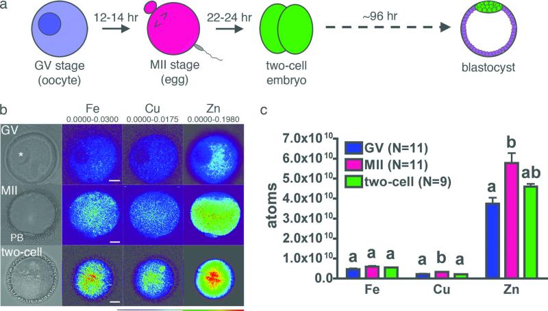
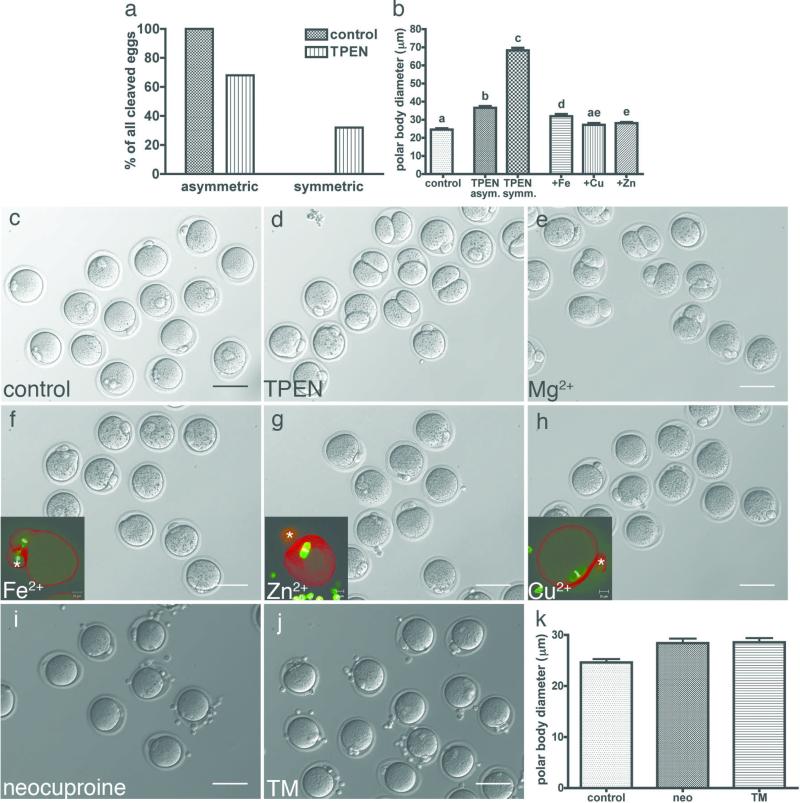
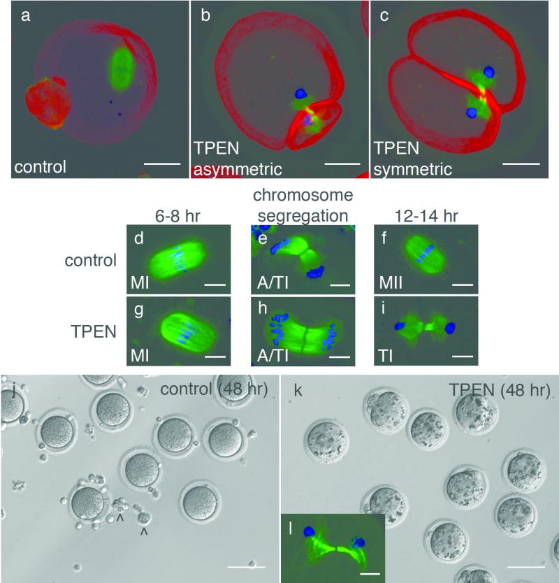
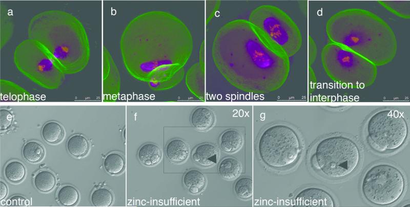
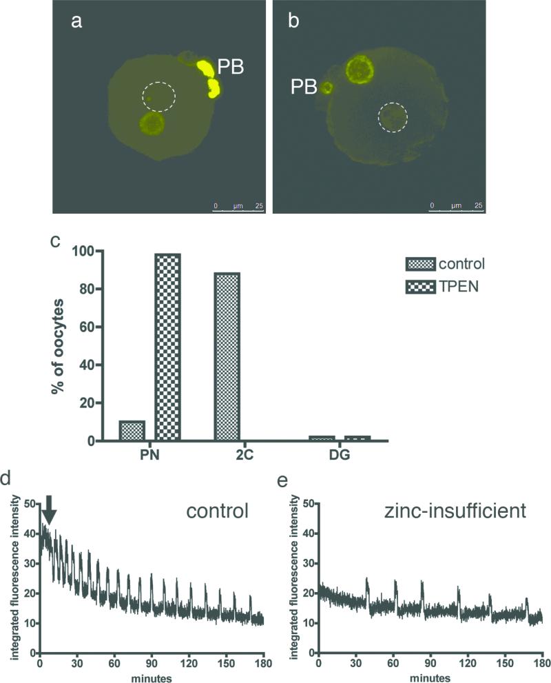
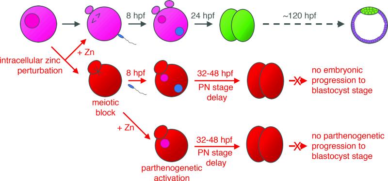
Cellular metal ion fluxes are known in alkali and alkaline earth metals but are not well documented in transition metals. Here we describe major changes in the zinc physiology of the mammalian oocyte as it matures and initiates embryonic development. Single-cell elemental analysis of mouse oocytes by synchrotron-based X-ray fluorescence microscopy (XFM) revealed a 50% increase in total zinc content within the 12-14-h period of meiotic maturation. Perturbation of zinc homeostasis with a cell-permeable small-molecule chelator blocked meiotic progression past telophase I. Zinc supplementation rescued this phenotype when administered before this meiotic block. However, after telophase arrest, zinc triggered parthenogenesis, suggesting that exit from this meiotic step is tightly regulated by the availability of a zinc-dependent signal. These results implicate the zinc bolus acquired during meiotic maturation as an important part of the maternal legacy to the embryo.


Figures

Figure 1
Figure 1. Synchrotron-based x-ray fluorescence microscopy reveals intracellular distribution of the transition elements in oocytes and early embryo
Fully-grown oocytes, eggs, and two-cell embryos (a) were prepared as whole mount samples for synchrotron-based xray fluorescence microscopy. Oocytes (N = 11) display an intact germinal vesicle (GV, asterisk) while mature (MII) eggs (N = 11) have a visible first polar body (PB). Two-cell embryos (N = 9) were obtained by in vitro fertilization. Representative brightfield images for each stage are shown, in addition to the elemental maps for iron, copper and zinc (b). The minimum and maximum elemental content (μg/cm2) are shown above each set of elements. Among the biologically relevant transition elements, zinc is an order of magnitude more abundant than iron and copper at all developmental stages (c). Scale bar = 20 μm. Data represent mean values ± s.e. Letters denote statistically significant differences between developmental stages for individual elements (p<0.05).
Figure 2
Figure 2. The heavy metal chelator TPEN disrupts asymmetric division of the oocyte, which can be rescued by exogenous zinc
Among those oocytes that extruded a polar body, 100% of control oocytes (N = 62) displayed asymmetric division (a). Only 68% of TPEN-treated oocytes (N = 65) displayed asymmetric division, while the remaining 32% were symmetrically divided. Measurement of the polar body diameter at its widest point showed that TPEN-treated oocytes had significantly larger polar bodies than control oocytes (b). Representative images of control (c) and TPEN-treated (d) oocytes are shown. Exogenous sources of magnesium (e), iron (f), zinc (g) and copper (h) were added directly to the chelator-containing medium after an initial maturation period of 4 hrs. Magnesium was included as a negative control as TPEN has low affinity for this metal. Copper (N = 34) and zinc (N = 71) supplementation restored polar body diameter more effectively than iron (N = 38) (b). Copper chelators neocuproine (i) and tetrathiomolybdate (TM, j) did not disrupt asymmetric division and polar body size remained indistinguishable from the control group (k). Scale bar = 80 μm. Data represent mean values ± s.e. Letters denote statistically significant differences in polar body diameter in b (p<0.05).
Figure 3
Figure 3. Zinc insufficient oocytes experience a meiotic block following telophase I
Control oocytes display a metaphase II spindle (a), but zinc-insufficient oocytes have a telophase I-like spindle with decondensed chromatin whether they cleave asymmetrically (b) or symmetrically (c). Control oocytes proceed through metaphase I (MI, d), chromosome segregation (anaphase/telophase I, or A/TI as shown in e), and establish a meiotic arrest at metaphase II (f). Zinc-insufficient oocytes also proceed through MI (g) and individual chromosomes are segregated to two poles (A/TI shown in h). However, by the time control oocytes reach MII, zinc-insufficient oocytes retain a telophase I spindle with decondensed chromatin at each pole (i). Control oocytes remain morphologically unchanged after a total of 48 hrs in culture (j). Zinc-insufficient oocytes become increasingly granular with extended culture (k) and they retain a telophase-like spindle with decondensed chromatin, although the tubulin array becomes less compact over time (l). Carats (^) denote cumulus cells. Scale bar = 20 μm (a-c), 10 μm (d-i, l), or 80 μm (j, k).
Figure 4
Figure 4. Zinc supplementation following telophase-like arrest induces spontaneous activation of the zinc-insufficient oocyte
Zinc supplementation at 12 hrs post-maturation is too late to restore the metaphase II spindle and a mixture of spindle configurations are seen (ad). After a full 16 hr maturation period, control (e) and zinc-insufficient (f) oocytes were transferred into KSOM for further culture. Control oocytes do not change over the extended culture period in the absence of an activating stimulus. In contrast, all zinc-insufficient oocytes (N = 62) exhibited pronuclear structures after 8 hrs (arrowheads, f and g). Magenta = alpha-tubulin, yellow = DNA, green = F-actin. Scale bar = 25 μm (a-d) or 80 μm (e, f).
Figure 5
Figure 5. Zinc-insufficient eggs can undergo a true fertilization event but display a delayed pronuclear stage and abnormal calcium oscillations upon activation
Fertilized control (a) and zinc-insufficient (b) eggs were fixed 8 hpf for 5-methylcytidine staining. In both groups, one of the two pronuclei displayed an absence of signal (dotted circles), indicating demethylation and a male origin of the genetic material. However, embryos derived from zinc-insufficient oocytes experience a delayed pronuclear stage (c). At 24 hpf, when embryos from control eggs have proceeded to the two-cell stage, embryos from zinc-insufficient eggs still display two pronuclei. Upon activation, control eggs initiate a prolonged first calcium transient (d, arrow) followed by a regular series of shorter transients. Zinc-insufficient oocytes lack a large first transient and undergo a fewer total number of transients within the same imaging period. PB = polar body, scale bar = 25 μm.
Figure 6
Figure 6. Summary of results
Normal maturation of fully-grown oocytes begins with the progression of an immature, germinal vesicle (GV) stage oocyte to a mature, metaphase II (MII)-arrested oocyte. Fertilization triggers the completion of meiosis and the first mitotic division results in the two-cell embryo, which will in turn develop into a blastocyst. Induction of zinc insufficiency during meiotic maturation results in oocytes that are prematurely arrested in meiotic telophase. Fertilization of these oocytes results in embryos that experience an extended pronuclear stage and impaired viability. This can be ameliorated with zinc supplementation prior to the establishment of telophase arrest. Simply returning zinc-insufficient oocytes to a zinc-replete environment triggers parthenogenetic activation in the absence of any other stimulus, leading to approximately two rounds of division before the parthenotes are no longer viable.

References

    1. Beyersmann D, Haase H. Functions of zinc in signaling, proliferation and differentiation of mammalian cells. Biometals. 2001;14:331–41. - PubMed
    1. Turski ML, Thiele DJ. New roles for copper metabolism in cell proliferation, signaling, and disease. J Biol Chem. 2009;284:717–21. - PMC - PubMed
    1. Zhang AS, Enns CA. Iron homeostasis: recently identified proteins provide insight into novel control mechanisms. J Biol Chem. 2009;284:711–5. - PMC - PubMed
    1. Yamasaki S, et al. Zinc is a novel intracellular second messenger. J Cell Biol. 2007;177:637–45. - PMC - PubMed
    1. Galaris D, Skiada V, Barbouti A. Redox signaling and cancer: the role of “labile” iron. Cancer Lett. 2008;266:21–9. - PubMed

Publication types

MeSH terms