. 2010 Sep;42(9):794-800.
doi: 10.1038/ng.641. Epub 2010 Aug 8.

Germline CBL mutations cause developmental abnormalities and predispose to juvenile myelomonocytic leukemia

Affiliations

Germline CBL mutations cause developmental abnormalities and predispose to juvenile myelomonocytic leukemia

Charlotte M Niemeyer et al. Nat Genet. 2010 Sep.

Abstract

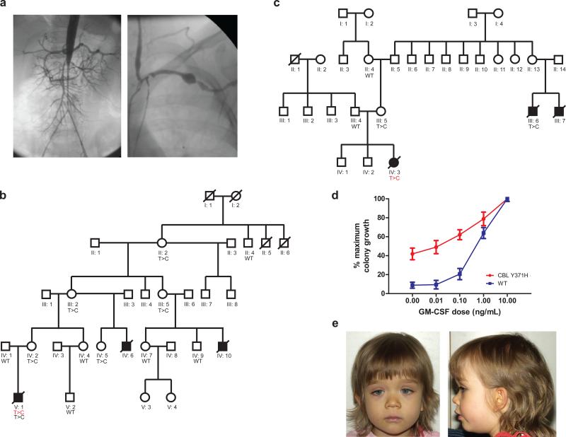
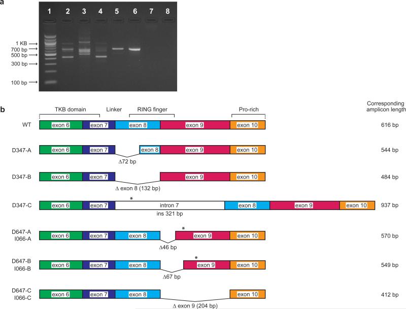
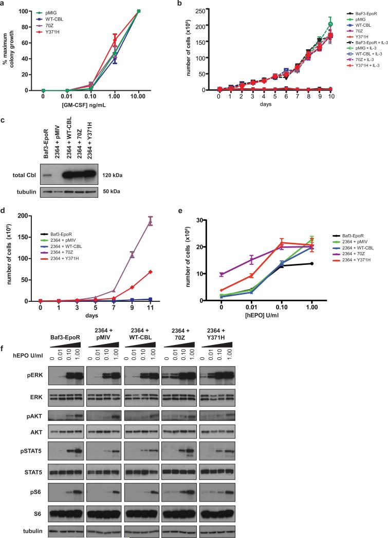
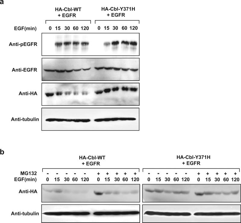
CBL encodes a member of the Cbl family of proteins, which functions as an E3 ubiquitin ligase. We describe a dominant developmental disorder resulting from germline missense CBL mutations, which is characterized by impaired growth, developmental delay, cryptorchidism and a predisposition to juvenile myelomonocytic leukemia (JMML). Some individuals experienced spontaneous regression of their JMML but developed vasculitis later in life. Importantly, JMML specimens from affected children show loss of the normal CBL allele through acquired isodisomy. Consistent with these genetic data, the common p.371Y>H altered Cbl protein induces cytokine-independent growth and constitutive phosphorylation of ERK, AKT and S6 only in hematopoietic cells in which normal Cbl expression is reduced by RNA interference. We conclude that germline CBL mutations have developmental, tumorigenic and functional consequences that resemble disorders that are caused by hyperactive Ras/Raf/MEK/ERK signaling and include neurofibromatosis type 1, Noonan syndrome, Costello syndrome, cardiofaciocutaneous syndrome and Legius syndrome.


Figures

Figure 1
Figure 1. Germline mutations in CBL can be inherited in an autosomal dominant fashion and are associated with a phenotype, GM-CSF hypersensitivity and vasculitis
Panel (a) demonstrates the angiograms from the aorta and left subclavian artery from patient D048 nine months after the diagnosis of Takayasu arteritis type III. Panel (b) The family tree of UPN1333 is shown in panel a, where the diseased bone marrow of UPN1333 displayed a homozygous CBL c.1111T>C (red) mutation as well as a heterozygous lesion from his buccal swab (black). Only women appear to be heterozygote carriers, and only boys appear to be affected by JMML in this family. Panel (c) The bone marrow of UPN1125 demonstrated a homozygous CBL mutation—her mother (III:5) is a known carrier, and she had two male cousins dying from JMML (III:6, III:7). Panel (d) demonstrates a classic GM-CSF hypersensitivity response on a colony assay for patients with CBL mutations (n=3) versus normal (n=13). Error bars represent standard error of the mean (s.e.m.) Panel (e) shows one toddler (D703) diagnosed with JMML and a homozygous mutation at p.C384R. She displays frontal bossing, downslanting palpebral fissues, hypertelorism, and a low nasal bridge. Photographs of her father, who harbors a heterozygous mutation at p.C384R, are included in Figures S1, panel d. Of note, both father and daughter also display bilateral ptosis.
Figure 2
Figure 2. Consequences of splice site mutations in cDNA from individuals D347, 647, and I066
Panel (a) RT-PCR using an exon 6 forward primer and an exon 10 reverse primer on cDNA generated from these patients. The wildtype amplicon is 616 base pairs long. Lane 1: MW ladder, Lane 2: I066, Lane 3: D347, Lane 4: D647, Lane 5: CBL point mutant, Lane 6: HM2833 CBL wildtype, Lane 7: Genomic DNA control, Lane 8: no template control. Panel (b) is a schematic representation of the splice site variants detected either recurrently (D347) or that were shared by I066 and D647. Sequences are shown in Figure S2. Deletions of base pairs are indicated by Δ# of base pairs and insertions by ins # base pairs. Premature stop codons are indicated by asterisks (*).
Figure 3
Figure 3. p.Y371H does not confer cytokine sensitivity or cytokine independent growth until silencing of murine Cbl
Panel (a) Transduction of p.Y371H or the known murine oncogeneic mutant 70Z in wildtype hematopoietic cells from fetal liver, did not confer hypersensitivity to GM-CSF, nor did expression of these mutants in BaF3-EpoR cells result in cytokine independent growth (b). Panel (c) An shRNA to murine Cbl (cbl.2364) demonstrated near complete shutdown of expression in BaF3-EpoR cells by Western blot with re-expression upon introduction of the human WT, 70Z, or p.Y371H in these same cell lines. Panel (d) Both the p.Y371H and 70Z Cbl conferred cytokine independent growth in the presence of cbl.2364. Controls included the Venus vector pMIV and BaF3-EpoR. Error bars for triplicate replicates (s.e.m.) are shown and when not visible, indicate tight clustering. Using a paired t-test: day 7 comparing 2364+ WT-Cbl versus 2364+p.Y371H, p-value= 0.017, and at day 9: p-value <0.001. Panel (e) Serial transduction of the hairpin (2364) and p.Y371H or 70Z constructs also conferred hypersensitive growth after assessing cell proliferation on day 5 in increasing concentrations of Epo. Using a paired t-test at each concentration of Epo when comparing 2364+WT-Cbl versus 2364+p.Y371H: Epo 0 unit/ml: p = 0.036, Epo 0.01 units/ml: p= 0.0015, Epo 0.1 units/ml: p= 0.029, Epo 1 unit/ml (saturating dose) P= 0.697. Panel (f) Both the p.Y371H and 70Z containing cells demonstrated activation of pERK, pAKT, and pS6 in the absence of Epo or in low dose 0.01 unit/mL of Epo in comparison to negative controls. All cell proliferation work was done in triplicate.
Figure 4
Figure 4. Cbl mutant proteins exhibit prolonged protein turnover and are associated with increased phosphorylated EGFR levels upon EGF stimulation
Panel (a) HEK293 cells transfected with plasmids encoding EGFR in combination with HA-Cbl(WT) or HA Cbl(p.Y371H) were serum starved for 18 h followed by 15 min EGF (50 ng/ml) stimulation. Cells were then washed and maintained in serum-free media for the indicated periods of time. Equal amounts of whole cell extracts were resolved on SDS-PAGE and immunoblotted with the indicated antibodies. Panel (b) HEK293 cells transfected with plasmids encoding EGFR in combination with HA-Cbl(WT) or HA-Cbl(p.Y371H) were serum starved for 18 h followed by 15 min EGF (50 ng/ml) stimulation. Cells were then washed and maintained in serum-free media with (+) or without (-) MG132 for the indicated periods of time. Equal amounts of whole cell extracts were resolved on SDS-PAGE and immunoblotted with the indicated antibodies.

References

    1. Niemeyer CM, Kratz CP. Paediatric myelodysplastic syndromes and juvenile myelomonocytic leukaemia: molecular classification and treatment options. Br J Haematol. 2008;140:610–24. - PubMed
    1. Locatelli F, et al. Hematopoietic stem cell transplantation (HSCT) in children with juvenile myelomonocytic leukemia (JMML): results of the EWOG-MDS/EBMT trial. Blood. 2005;105:410–9. - PubMed
    1. Matsuda K, et al. Spontaneous improvement of hematologic abnormalities in patients having juvenile myelomonocytic leukemia with specific RAS mutations. Blood. 2007;109:5477–80. - PubMed
    1. Flotho C, et al. Genotype-phenotype correlation in cases of juvenile myelomonocytic leukemia with clonal RAS mutations. Blood. 2008;111:966–7. author reply 967-8. - PubMed
    1. Niemeyer CM, et al. Chronic myelomonocytic leukemia in childhood: a retrospective analysis of 110 cases. European Working Group on Myelodysplastic Syndromes in Childhood (EWOG-MDS). Blood. 1997;89:3534–43. - PubMed

Publication types

Substances