

Inhibitory actions of mibefradil on steroidogenesis in mouse Leydig cells: involvement of Ca(2+) entry via the T-type Ca(2+) channel
- PMID: 20694017
- PMCID: PMC3739071
- DOI: 10.1038/aja.2010.51
Inhibitory actions of mibefradil on steroidogenesis in mouse Leydig cells: involvement of Ca(2+) entry via the T-type Ca(2+) channel
Abstract
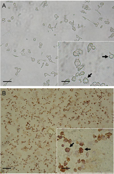
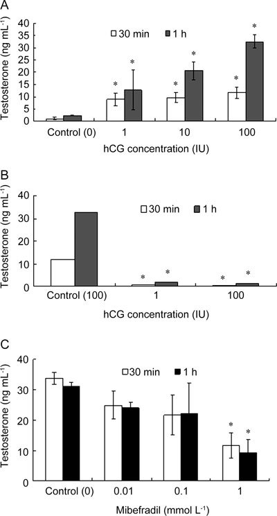
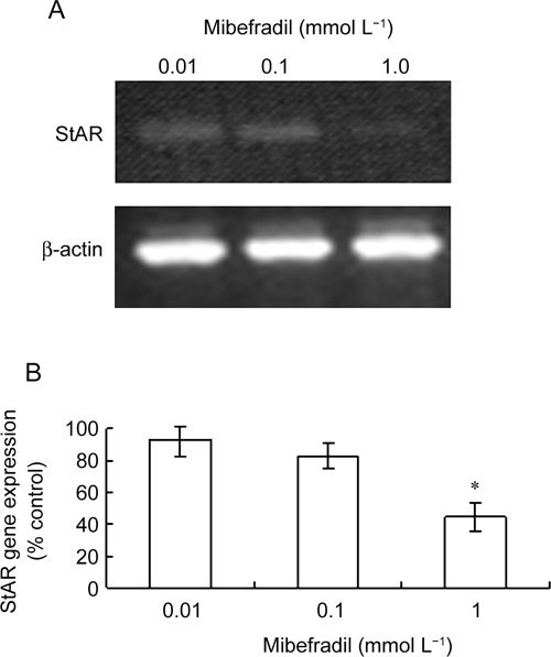
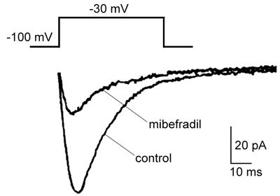
Intracellular cAMP and Ca(2+) are involved in the regulation of steroidogenic activity in Leydig cells, which coordinate responses to luteinizing hormone (LH) and human chorionic gonadotropin (hCG). However, the identification of Ca(2+) entry implicated in Leydig cell steroidogenesis is not well defined. The objective of this study was to identify the type of Ca(2+) channel that affects Leydig cell steroidogenesis. In vitro steroidogenesis in the freshly dissociated Leydig cells of mice was induced by hCG incubation. The effects of mibefradil (a putative T-type Ca(2+) channel blocker) on steroidogenesis were assessed using reverse transcription (RT)-polymerase chain reaction analysis for the steroidogenic acute regulatory protein (StAR) mRNA expression and testosterone production using radioimmunoassay. In the presence of 1.0 mmol L(-1) extracellular Ca(2+), hCG at 1 to 100 IU noticeably elevated both StAR mRNA level and testosterone secretion (P < 0.05), and the stimulatory effects of hCG were markedly diminished by mibefradil in a dose-dependent manner (P < 0.05). Moreover, the hCG-induced increase in testosterone production was completely removed when external Ca(2+) was omitted, implying that Ca(2+) entry is needed for hCG-induced steroidogenesis. Furthermore, a patch-clamp study revealed the presence of mibefradil-sensitive Ca(2+) currents seen at a concentration range that nearly paralleled those inhibiting steroidogenesis. Collectively, our data provide evidence that hCG-stimulated steroidogenesis is mediated at least in part by Ca(2+) entry carried out by the T-type Ca(2+) channel in the Leydig cells of mice.
Figures
Similar articles
-
Functional assessment of the calcium messenger system in cultured mouse Leydig tumor cells: regulation of human chorionic gonadotropin-induced expression of the steroidogenic acute regulatory protein.Endocrinology. 1999 Apr;140(4):1739-51. doi: 10.1210/endo.140.4.6650. Endocrinology. 1999. PMID: 10098511
-
Blocking L-type calcium channels reduced the threshold of cAMP-induced steroidogenic acute regulatory gene expression in MA-10 mouse Leydig cells.J Endocrinol. 2010 Jan;204(1):67-74. doi: 10.1677/JOE-09-0206. Epub 2009 Oct 12. J Endocrinol. 2010. PMID: 19822634 Free PMC article.
-
Effects of genistein, resveratrol, and quercetin on steroidogenesis and proliferation of MA-10 mouse Leydig tumor cells.J Endocrinol. 2007 Mar;192(3):527-37. doi: 10.1677/JOE-06-0087. J Endocrinol. 2007. PMID: 17332522
-
T-Type Calcium Channels: A Mixed Blessing.Int J Mol Sci. 2022 Aug 31;23(17):9894. doi: 10.3390/ijms23179894. Int J Mol Sci. 2022. PMID: 36077291 Free PMC article. Review.
-
Knowledge Gap in Understanding the Steroidogenic Acute Regulatory Protein Regulation in Steroidogenesis Following Exposure to Bisphenol A and Its Analogues.Biomedicines. 2022 May 30;10(6):1281. doi: 10.3390/biomedicines10061281. Biomedicines. 2022. PMID: 35740303 Free PMC article. Review.
Cited by
-
Rapid responses to reverse T₃ hormone in immature rat Sertoli cells: calcium uptake and exocytosis mediated by integrin.PLoS One. 2013 Oct 10;8(10):e77176. doi: 10.1371/journal.pone.0077176. eCollection 2013. PLoS One. 2013. PMID: 24130850 Free PMC article.
-
Gonadotropin-releasing hormone positively regulates steroidogenesis via extracellular signal-regulated kinase in rat Leydig cells.Asian J Androl. 2011 May;13(3):438-45. doi: 10.1038/aja.2010.158. Epub 2011 Mar 28. Asian J Androl. 2011. PMID: 21441942 Free PMC article.
-
Calcium Signaling in Interstitial Cells: Focus on Telocytes.Int J Mol Sci. 2017 Feb 13;18(2):397. doi: 10.3390/ijms18020397. Int J Mol Sci. 2017. PMID: 28208829 Free PMC article. Review.
-
Regulation of testosterone synthesis in Leydig cells by ClC-2 chloride channel.Reproduction. 2025 Jul 18;170(2):e240432. doi: 10.1530/REP-24-0432. Print 2025 Aug 1. Reproduction. 2025. PMID: 40683311 Free PMC article.
References
-
- Dufau ML. Endocrine regulation and communicating functions of the Leydig cell. Ann Rev Physiol. 1988;50:483–508. - PubMed
-
- Saez JM. Leydig cells: endocrine, paracrine, and autocrine regulation. Endocr Rev. 1994;15:574–626. - PubMed
-
- Chen YC, Nagpal ML, Stocco DM, Lin T. Effects of genistein, resveratrol, and quercetin on steroidogenesis and proliferation of MA-10 mouse Leydig tumor cells. J Endocrinol. 2007;192:527–37. - PubMed
-
- Medelson C, Dufau ML, Catt KJ. Gonadotropin binding and stimulation of cAMP and testosterone production in isolated Leydig cells. J Biol Chem. 1975;250:8818–23. - PubMed
Publication types
MeSH terms
Substances
LinkOut - more resources
Full Text Sources
Miscellaneous
