. 2010 Aug 4;5(8):e11953.
doi: 10.1371/journal.pone.0011953.

Genome-wide analysis of effectors of peroxisome biogenesis

Affiliations

Genome-wide analysis of effectors of peroxisome biogenesis

Ramsey A Saleem et al. PLoS One. .

Abstract

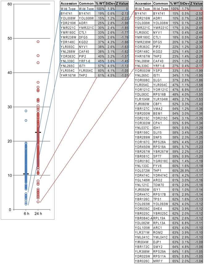
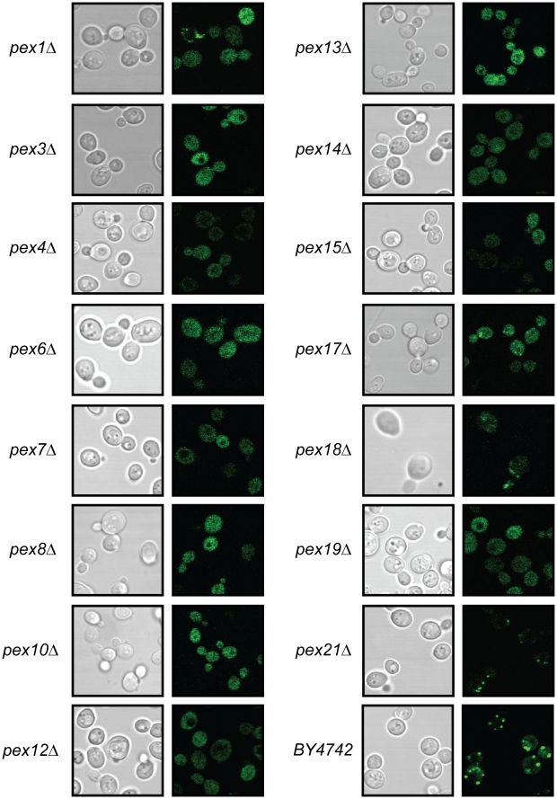
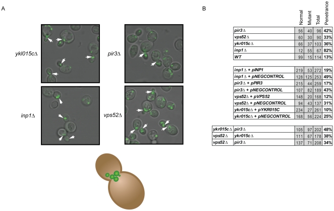
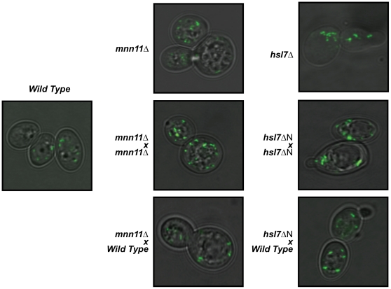
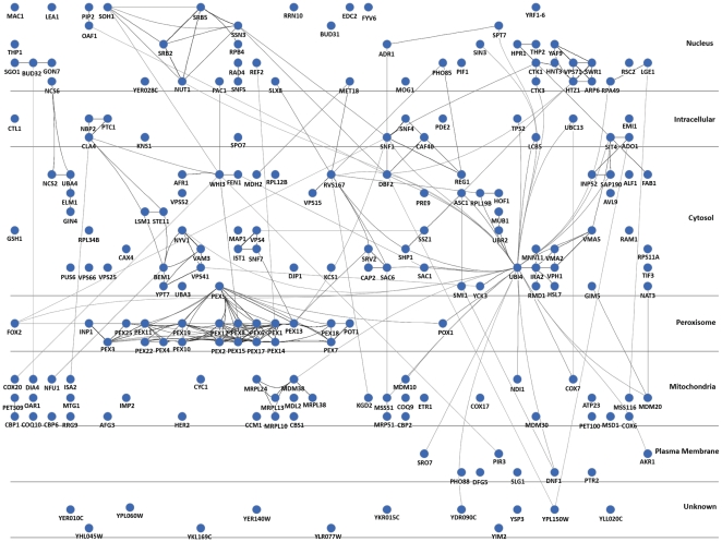
Peroxisomes are intracellular organelles that house a number of diverse metabolic processes, notably those required for beta-oxidation of fatty acids. Peroxisomes biogenesis can be induced by the presence of peroxisome proliferators, including fatty acids, which activate complex cellular programs that underlie the induction process. Here, we used multi-parameter quantitative phenotype analyses of an arrayed mutant collection of yeast cells induced to proliferate peroxisomes, to establish a comprehensive inventory of genes required for peroxisome induction and function. The assays employed include growth in the presence of fatty acids, and confocal imaging and flow cytometry through the induction process. In addition to the classical phenotypes associated with loss of peroxisomal functions, these studies identified 169 genes required for robust signaling, transcription, normal peroxisomal development and morphologies, and transmission of peroxisomes to daughter cells. These gene products are localized throughout the cell, and many have indirect connections to peroxisome function. By integration with extant data sets, we present a total of 211 genes linked to peroxisome biogenesis and highlight the complex networks through which information flows during peroxisome biogenesis and function.


Conflict of interest statement

Competing Interests: The authors have declared that no competing interests exist.

Figures

Figure 1
Figure 1. Flow cytometry analysis of candidate deletion strains.
Plot of the log10 fluorescence of candidate deletion strains tested at 6 h and 24 h of oleate incubation. Deletion strains are indicated with open circles, while wild type is indicated by the line. The tables at the right show the flow cytometry analysis of the candidate genes with percentage of Pot1p-GFP fluorescence relative to wild type, the mean of the fluorescence values and the z value or standard score of the fluorescence at 6 h or 24 h respectively. Genes are shown that show decreases of Pot1p-GFP fluorescence that are at least 1SD from the wild type POT1-GFP strain. Genes boxed by blue or red indicate the naturally occurring separation of values shown in the plot at 6 h (blue) and 24 h (red). The remaining flow cytometry results are shown in Table S1-2. BY4741 is used as a non-fluorescent control strain.
Figure 2
Figure 2. Identification of peroxisomal mutants.
Bright field images are shown on the left panel and fluorescence images are shown on the right. Known peroxins show mislocalization of the Pot1p-GFP reporter when deleted. Partial mislocalization phenotypes are seen in pex1Δ, pex18Δ, and pex21Δ.
Figure 3
Figure 3. Vps52p, Pir3p and YKL015C are novel peroxisome inheritance factors.
A. Strains lacking Inp1p, Vps52p, Pir3p or YKL015Cp accumulate peroxisomes near the bud neck or in daughter cells as indicated by the arrowheads. B. Summary of the effect of (i) deletions, (ii) complementation, and (iii) double deletions of vps52Δ, pir3Δ and ykl015cΔ.
Figure 4
Figure 4. Mnn11p and Hsl7p are regulators of peroxisome morphology.
Strains deleted for mnn11 or the 5′ end of hsl7 (hsl7ΔN generated by the ybr134w deletion) were backcrossed against an allelic deletion or a wild type strain (BY4742). An increased number of small peroxisomes are seen in cells deleted for mnn11, while hsl7ΔN lead to an increase in size or tendency for the peroxisomes to cluster. Both phenotypes are complemented by mating to the wild type strain.
Figure 5
Figure 5. The cellular network regulating biogenesis of peroxisomes and the utilization of fatty acids.
The circular nodes represent proteins identified in Table S2 and protein-protein interactions from the literature shown by edges between the nodes. For ease of display, protein localized to the vacuole, ER and Golgi are represented by the annotation “Cytosol”. Distinct functional modules within the nucleus, peroxisomal and mitchondrial localizations can be seen as well as the high connectivity of ubiquitin (UBI4).

Similar articles

Cited by

References

    1. Smith JJ, Aitchison JD. Regulation of peroxisome dynamics. Curr Opin Cell Biol. 2009;21:119–126. - PMC - PubMed
    1. Saleem RA, Knoblach B, Mast FD, Smith JJ, Boyle J, et al. Genome-wide analysis of signaling networks regulating fatty acid-induced gene expression and organelle biogenesis. J Cell Biol. 2008;181:281–292. - PMC - PubMed
    1. Wan Y, Saleem RA, Ratushny AV, Roda O, Smith JJ, et al. Role of the histone variant H2A.Z/Htz1p in TBP recruitment, chromatin dynamics, and regulated expression of oleate-responsive genes. Mol Cell Biol. 2009;29:2346–2358. - PMC - PubMed
    1. Koerkamp MG, Rep M, Bussemaker HJ, Hardy GP, Mul A, et al. Dissection of transient oxidative stress response in Saccharomyces cerevisiae by using DNA microarrays. Mol Biol Cell. 2002;13:2783–2794. - PMC - PubMed
    1. Smith JJ, Marelli M, Christmas RH, Vizeacoumar FJ, Dilworth DJ, et al. Transcriptome profiling to identify genes involved in peroxisome assembly and function. J Cell Biol. 2002;158:259–271. - PMC - PubMed

Publication types

Substances