. 2010 Oct;6(10):2049-55.
doi: 10.1039/c004684a. Epub 2010 Aug 9.

Lipo-oligoarginines as effective delivery vectors to promote cellular uptake

Affiliations

Lipo-oligoarginines as effective delivery vectors to promote cellular uptake

Jae Sam Lee et al. Mol Biosyst. 2010 Oct.

Abstract

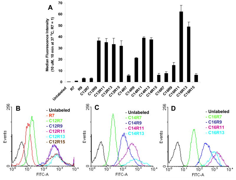
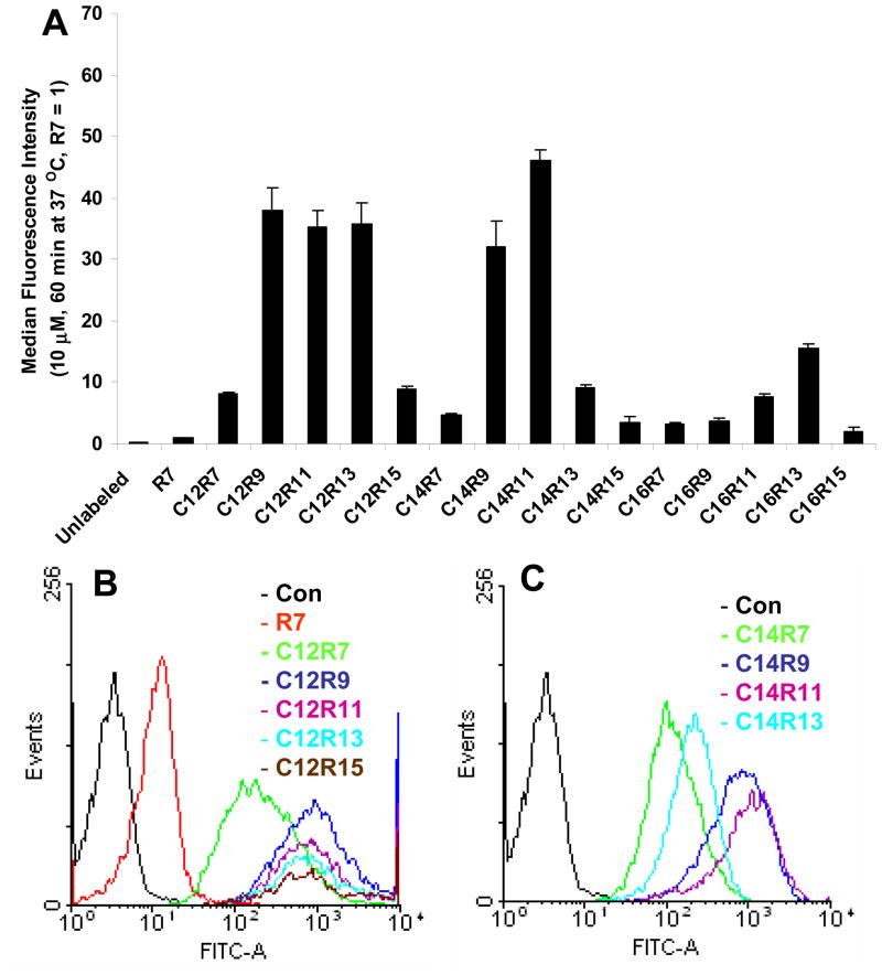
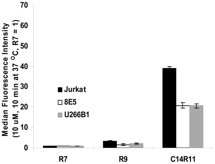
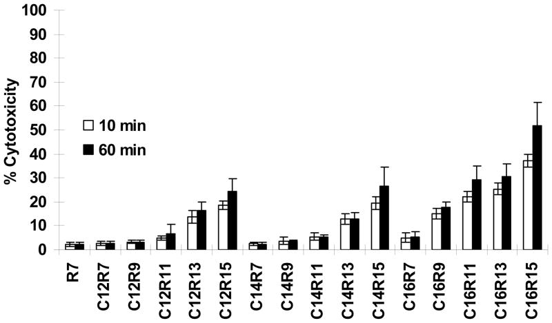
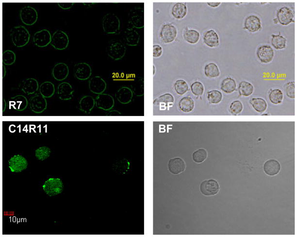
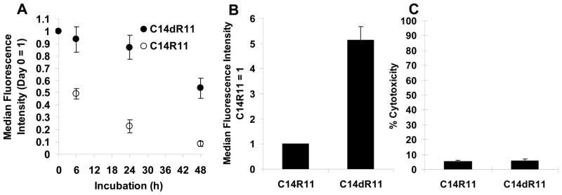
An effective cellular delivery vector with enhanced intracellular retention was developed by conjugating a cell-penetrating peptide (CPP) with a fatty acid chain. The optimized lipopeptide (LP), myristoylated hendecaarginine (C14R11), penetrated the cell membrane with high efficiency, and achieved superior metabolic stability and versatility as compared with unmodified oligoarginine CPPs, offering no adverse effect on cell viability and function. Cellular uptake, intracellular localization, cytotoxicity, and release kinetics of oligoarginines and LPs were investigated using flow cytometry analysis, cytotoxicity assay, and confocal microscopy. The cellular uptake efficiency and intracellular metabolic stability of C14R11 LP was further enhanced by replacing the L-arginine residues with D-arginine isomers. The cellular uptake and intracellular metabolic stability of D-form C14R11 (C14dR11) was significantly increased without any noticeable cytotoxicity compared to the unmodified parent hepta-arginine CPP or L-arginine LPs.


Figures

Fig. 1
Fig. 1
Cellular uptake of 10 μM LPs incubated with Jurkat cells for 10 min at 37 °C. (A) Jurkat cells incubated with LPs were washed, trypsin treated, PI stained, and analyzed by flow cytometry with 1–2 × 10⁴ events. (B, C, and D) The histograms of C12, C14, or C16 LPs. Each experiment was performed at least three times in triplicate, and the results are expressed as the median change in fluorescence ± S.D. The median fluorescence intensity of R7 in Jurkat cell was set as 1.
Fig. 2
Fig. 2
Cellular uptake of LPs in Jurkat cells incubated for 60 min. (A) Cells are incubated with 10 μM of LPs for 60 min at 37 °C. The fluorescence intensity of R7 at 60 min was set as 1. (B and C) The flow cytometry histograms of C12 and C14 LPs.
Fig. 3
Fig. 3
Cellular uptake of 10 μM LPs incubated with various cell lines. Jurkat, 8E5, and U266B1 cells were incubated with R7, R9, and C14R11 LPs, washed, and analyzed by flow cytometry. The median fluorescence intensity of R7 in each cell line was set as 1.
Fig. 4
Fig. 4
Effects of LPs on cell toxicity. Jurkat cells were incubated with 10μM of LPs for 10 min or 60 min at 37 °C. Data were normalized to the amount of maximum LDH released from the cultures treated with lysis solution alone and corrected for baseline LDH release from cultures exposed to buffer only.
Fig. 5
Fig. 5
Cellular uptake of LPs at different temperatures. (A) Relative fluorescent intensities of LPs. Cells were incubated with 10 μM of LPs (R7, C12R9, or C14R11) at 4 °C for 10–60 min. The fluorescence intensity of R7 incubated for 10 min at 4 °C was set as 1. (B) Absolute fluorescent intensity of cells at 4 °C or 37 °C for 10 or 60 min.
Fig. 6
Fig. 6
Intracellular distribution of LPs in Jurkat cells. Cells were treated with 10 μM of R7 and C14R11 for 10 min at 37 °C. The cell images were acquired by confocal microscopy.
Fig. 7
Fig. 7
Comparison of C14dR11 or C14R11 on Jurkat cells. (A) Retention profiles of LPs. Cells were cultured with 10 μM of C14R11 or C14dR11 LPs at 37 °C for 10 min. Then cells were washed and reincubated in 6 well culture plates for a period of time (0, 6, 24, and 48 h) prior to flow cytometry analysis. (B) Cellular uptake of C14R11 or C14dR11 LPs. Cells was incubated with 10 μM of LPs at 37 °C for 10 min. The fluorescence intensity of C14R11 peptide was set as 1. (C) Cytotoxicity of C14R11 and C14dR11 LPs. Cells were incubated with 10 μM of LPs at 37 °C for 60 min. Experiments were performed in triplicate, mean ± SD.

References

    1. Frankel AD, Pabo CO. Cell. 1988;55:1189–1193. - PubMed
    1. Green M, Loewenstein PM. Cell. 1988;55:1179–1188. - PubMed
    1. Joliot AH, Triller A, Volovitch M, Pernelle C, Prochiantz A. New Biol. 1991;3:1121–1134. - PubMed
    1. Derossi D, Calvet S, Trembleau A, Brunissen A, Chassaing G, Prochiantz A. J Biol Chem. 1996;271:18188–18193. - PubMed
    1. Vives E, Brodin P, Lebleu B. J Biol Chem. 1997;272:16010–16017. - PubMed

Publication types