. 2011 Mar;469(3):860-7.
doi: 10.1007/s11999-010-1497-z. Epub 2010 Aug 6.

Application of the Ilizarov technique to the correction of neurologic equinocavovarus foot deformity

Affiliations

Application of the Ilizarov technique to the correction of neurologic equinocavovarus foot deformity

Dong Yeon Lee et al. Clin Orthop Relat Res. 2011 Mar.

Abstract

Background: The treatment of rigid equinocavovarus foot deformities caused by neurologic disorders is often difficult and relapse is common.

Questions/purposes: We asked whether the Ilizarov technique could be used for correction of neurologic equinocavovarus foot deformities resulting in improved foot and ankle function and patient satisfaction.

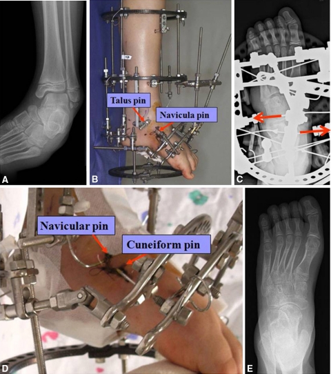
Patients and methods: The neurologic equinocavovarus foot deformities of 26 patients (mean age, 18.7 years; 29 feet) were treated using the Ilizarov technique. Nine feet were treated by distraction histiogenesis only with limited soft tissue release, whereas 20 feet needed additional osteotomy and/or tendon transfer/lengthening. Minimum followup was 12 months (mean, 72.9 months; range, 12-155 months).

Results: The mean time required for deformity correction was 27.1 days (range, 14-47 days) and the mean time for stabilization in the apparatus was 23.2 days (range, 7-53 days). A painless, stable, and plantigrade result was obtained by 22 patients (24 feet). Mild residual foot deformity was observed in the remaining five feet of four patients. Six patients (six feet) experienced postoperative complications. Three patients (four feet) experienced recurrence of the deformity requiring surgical correction.

Conclusions: Ilizarov soft tissue distraction with or without callotasis of tarsal bone(s) allows a greater degree of correction of neurologic equinocavovarus foot deformities. However, to reduce the risk of recurrence after fixator removal, it may be necessary to overcorrect the deformity while in the fixator, to use nighttime splinting, and most importantly, to eliminate neuromuscular imbalance, if necessary, by combining arthrodesis with or without tendon transfer.

Level of evidence: Level IV, therapeutic study. See Guidelines for Authors for a complete description of levels of evidence.


Figures

Fig. 1A–E
Fig. 1A–E
(A) Talonavicular joint subluxation is best seen in this projection of the plain radiograph. (B) A transfixing olive pin is inserted from the lateral to medial direction in the talar neck to pull the externally rotated talar head medialward. Then, another transfixing olive pin is inserted from the medial to lateral direction in the navicula, which is attached to the forefoot ring. (C) The navicular olive pin therefore provides a counterforce against the talar pin while reducing the talonavicular joint by pulling the medial rod to rotate the talus medially. (D) After reducing the talonavicular joint, the navicula stirrup wire position is transposed to the tibial ring to allow distraction osteogenesis of the plantar-medial aspect of the cuneiform between the midfoot and forefoot. (E) Talonavicular joint reduction and good hindfoot alignment were achieved.
Fig. 2A–H
Fig. 2A–H
The photographs show representative examples of neurologic equinocavovarus deformity according to the modified Dimeglio classification (Table 2). (A) Anteroposterior and (B) posteroanterior photographs show a severe deformity with a total score of 9. (C) Anteroposterior and (D) posteroanterior photographs show a moderate deformity with a total score of 5. These (E) anteroposterior and (F) posteroanterior photographs show a mild deformity with a total score of 2, and these (G) anteroposterior and (H) posteroanterior photographs show a postural foot with a total score of 0.
Fig. 3A–G
Fig. 3A–G
A 10-year-old boy with spastic cerebral palsy, hemiplegic type, who had no previous operations, underwent correction of his deformity using the Ilizarov technique. (A) Anteroposterior and (B) posteroanterior photographs and (C) a preoperative lateral radiograph show his severe equinocavovarus foot deformity. (D) A postoperative photograph taken just before removal of the Ilizarov fixator shows nice correction of the foot deformity. (E) Anterioposterior and (F) posteroanterior photographs and (G) a radiograph taken 4 years after surgery show excellent results with postural plantigrade feet.
Fig. 4
Fig. 4
A flowchart shows an algorithmic approach in decision-making for the treatment of neurologic equinocavovarus foot deformities using Ilizarov methods.

Similar articles

Cited by

References

    1. Atar D, Lehman WB, Grant AD. Complications in clubfoot surgery. Orthop Rev. 1991;20:233–239. - PubMed
    1. Carroll NC. Controversies in the surgical management of clubfoot. Instr Course Lect. 1996;45:331–337. - PubMed
    1. Carroll NC, McMurtry R, Leete SF. The pathoanatomy of congenital clubfoot. Orthop Clin North Am. 1978;9:225–232. - PubMed
    1. Choi IH, Kim JI, Yoo WJ, Chung CY, Cho TJ. Ilizarov treatment for equinoplanovalgus foot deformity caused by melorheostosis. Clin Orthop Relat Res. 2003;414:238–241. doi: 10.1097/01.blo.0000076801.53006.80. - DOI - PubMed
    1. Choi IH, Yang MS, Chung CY, Cho TJ, Sohn YJ. The treatment of recurrent arthrogrypotic club foot in children by the Ilizarov method: a preliminary report. J Bone Joint Surg Br. 2001;83:731–737. doi: 10.1302/0301-620X.83B5.11019. - DOI - PubMed