Case Reports
. 2010 Nov;39(11):1145-9.
doi: 10.1007/s00256-010-1014-1. Epub 2010 Aug 8.

Osteoid osteoma of the femur in a 7-month-old infant treated with radiofrequency ablation

Affiliations
Case Reports

Osteoid osteoma of the femur in a 7-month-old infant treated with radiofrequency ablation

Warapat Virayavanich et al. Skeletal Radiol. 2010 Nov.

Abstract

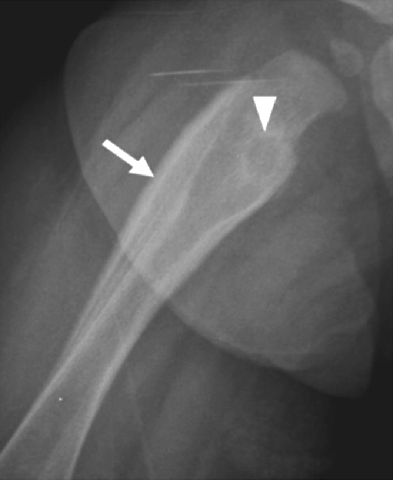
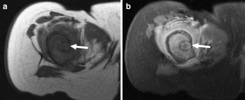
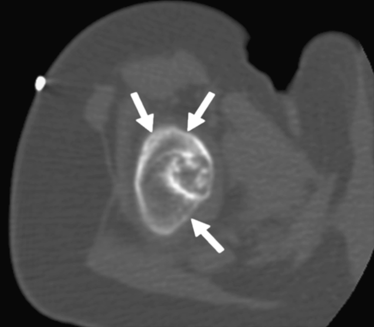
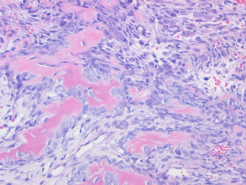
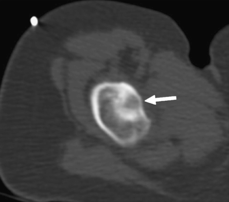
Osteoid osteoma occurs most commonly in children, adolescents, and young adults between the ages of 5 and 30 years. In the preschool age group, it is quite uncommon, accounting for only 3-8% of all osteoid osteoma cases. We report a case of osteoid osteoma in a 7-month-old infant, who presented with decreased use of the right lower extremity due to pain. Magnetic resonance imaging (MRI) showed an atypical appearance. A biopsy of the lesion, with histopathological examination, confirmed the diagnosis of osteoid osteoma. Radiofrequency ablation (RFA) of the nidus under computed tomography (CT) guidance was performed. The patient developed a recurrence after 3 months, which was treated with a second RFA. On subsequent follow-up, the infant did not show signs of pain after 1 month. In summary, this case report shows that osteoid osteoma can present in early infancy and can be successfully treated with RFA at this age, however, recurrence after the procedure can occur and close follow-up is recommended.


Figures

Fig. 1
Fig. 1
Roentgenogram of the right hip shows a lytic lesion with sclerotic rim centered in the intertrochanteric region of the proximal right femur (arrowhead). There is lamellar periosteal reaction involving the proximal femur extending to the mid diaphysis (arrow)
Fig. 2
Fig. 2
Axial T1-weighted pre-contrast (a) and fat-saturated post-contrast (b) MR images of the right femur show a tumor nidus (arrows) associated with extensive bone marrow edema pattern and extra-osseous edema pattern involving the proximal metadiaphysis and diaphysis
Fig. 3
Fig. 3
Axial CT scan (bone window) during percutaneous RFA shows a radiolucent intramedullary nidus with central calcification surrounded by a dense rim of sclerosis. There is a marked periosteal reaction around the cortex (arrows). Note a marker for planning the skin entry point
Fig. 4
Fig. 4
The tumor consisted of delicate trabeculae of woven bone with osteoblastic rimming. The intertrabecular space was occupied by a loose, hypocellular, vascular stroma. (Hematoxylin and eosin stain, 400× original magnification)
Fig. 5
Fig. 5
Axial CT scan (bone window) obtained during the RFA of the recurrent OO. Note mild decrease in size of the nidus (arrow) and periosteal bone formation

Similar articles

Cited by

References

    1. Dorfman HD, Czerniak B. Benign osteoblastic tumors. In: Dorfman HD, Czerniak B, editors. Bone tumors. St. Louis: Mosby; 1998. pp. 85–127.
    1. Cohen MD, Harrington TM, Ginsburg WW. Osteoid osteoma: 95 cases and a review of the literature. Semin Arthritis Rheum. 1983;12:265–281. doi: 10.1016/0049-0172(83)90010-0. - DOI - PubMed
    1. Jackson RP, Reckling FW, Mants FA. Osteoid osteoma and osteoblastoma. Similar histologic lesions with different natural histories. Clin Orthop Relat Res. 1977;128:303–313. - PubMed
    1. Schajowicz F, Lemos C. Osteoid osteoma and osteoblastoma. Closely related entities of osteoblastic derivation. Acta Orthop Scand. 1970;41:272–291. doi: 10.3109/17453677008991514. - DOI - PubMed
    1. Habermann ET, Stern RE. Osteoid-osteoma of the tibia in an eight-month-old boy. A case report. J Bone Joint Surg Am. 1974;56:633–636. - PubMed

Publication types

MeSH terms