Review
. 2010 Dec;67(23):4001-9.
doi: 10.1007/s00018-010-0479-3. Epub 2010 Aug 8.

Cellular communications in bone homeostasis and repair

Affiliations
Review

Cellular communications in bone homeostasis and repair

Ken-Ichi Nakahama. Cell Mol Life Sci. 2010 Dec.

Abstract

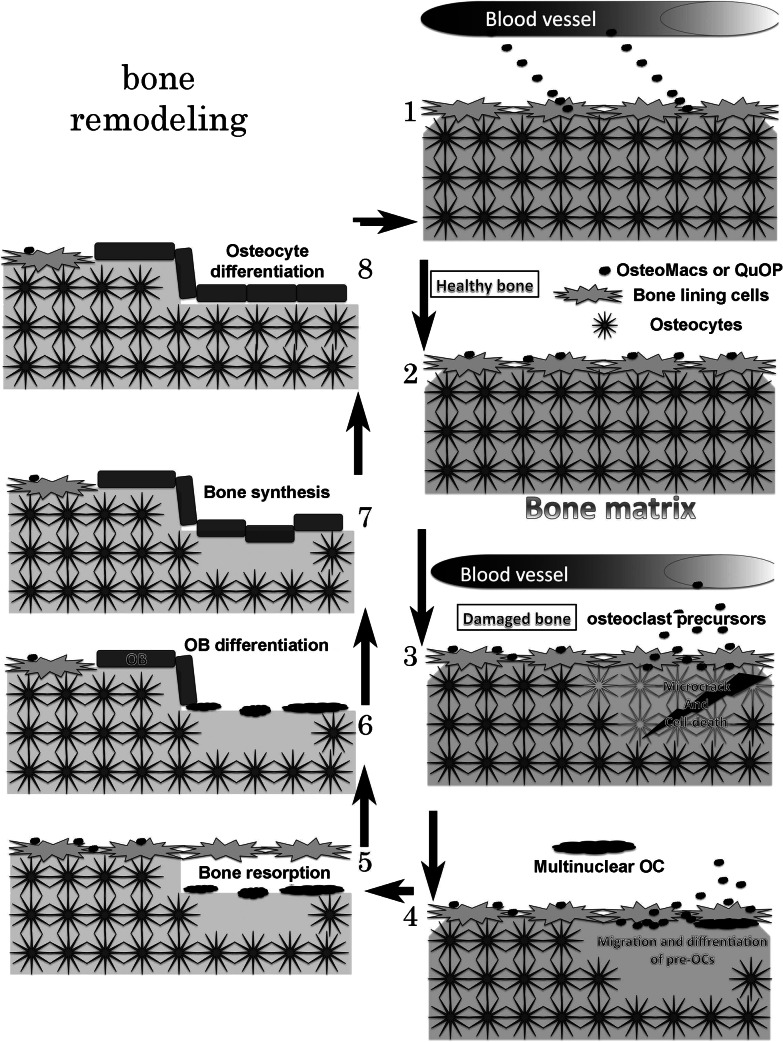
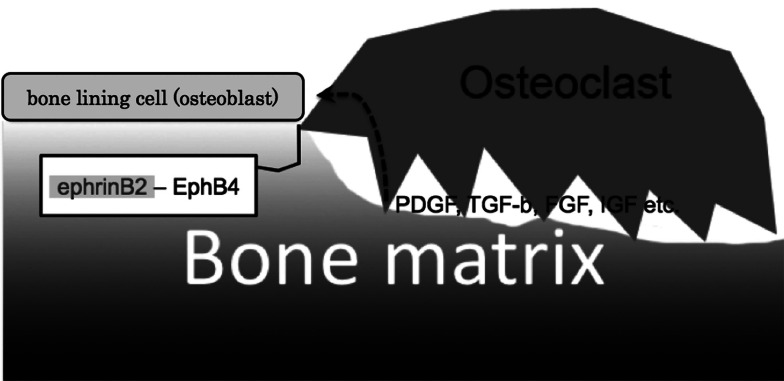
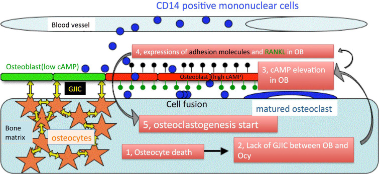
Cellular communication between the bone component cells osteoblasts, osteocytes and (pre-)osteoclasts is essential for bone remodeling which maintains bone integrity. As in the remodeling of other organs, cell death is a trigger for remodeling of bone. During the systematic process of bone remodeling, direct or indirect cell-cell communication is indispensable. Thus, osteoblasts induce migration and differentiation of preosteoclasts, which is followed by bone resorption (by mature multinuclear osteoclasts). After completion of bone resorption, apoptosis of mature osteoclasts and differentiation of osteoblasts are initiated. At this time, the osteoblasts do not support osteoclast differentiation but do support bone formation. Finally, osteoblasts differentiate to osteocytes in bone or to bone lining cells on bone surfaces. In this way, old bone areas are regenerated as new bone. In this review the role of cell-cell communication in bone remodeling is discussed.


Figures

Fig. 1
Fig. 1
Schematic illustration of bone remodeling. 1 Bone marrow-derived mononuclear cells (BMMs, osteoclast precursors) migrate on osteoblast. 2 In healthy bone, osteoclast precursors stay on the osteoblast layer and are named osteal macrophages (OsteoMacs) or cell cycle-arrested quiescent osteoclast precursors (QuOP). 3 A microcrack in the bone induces osteocyte apoptosis following which, osteal (or QuOP) and BM-derived macrophages migrate under the osteoblast layer. The macrophages may differentiate into preosteoclast (pre-OC). 4 The pre-OCs fuse with each other under the osteoblast layer. 5 Multinuclear osteoclasts erode the bone matrix containing dead osteocytes. 6 Osteogenic osteoblasts proliferate and cover the bone resorption area. 7 Bone is synthesized. 8 Some osteoblasts differentiate into osteocytes
Fig. 2
Fig. 2
Confocal microscopic images of the calvaria of an adult, CAG promoter-driven green fluorescence protein transgenic mouse. Optical sections (x, y) are shown with additional xz and yz projections. Osteocyte–osteocyte and osteoblast–osteocyte communications were found. Bar 10 μm. a Image focused on an osteocyte (asterisks). b Image focused on the osteoblast layer (hashes)
Fig. 3
Fig. 3
Cell communication between MNCs and endothelial cells. MNCs migrate from the bloodstream to the extravascular space. Extravasation of MNCs is controlled by adhesion molecules (ICAM-1 and LFA-1, CD44). Two migratory routes through the endothelia have been reported (paracellular and transcellular migration)
Fig. 4
Fig. 4
Cell communication between osteoclast precursors (MNCs) and osteoblasts. MNCs migrate under osteoblasts. This migration is also mediated by adhesion molecules (ICAM-1-Mac1, ICAM-1-LFA-1, CX3CR1-CX3CL1). Two migratory routes have been proposed (paracellular and transcellular migration). After MNCs have migrated under the osteoblast layer, they differentiate into pre-OCs, which are induced by RANK-RANKL binding. Osteoblasts express ICAM-1, CX3CR1 and RANKL. Pre-OCs express Mac-1, LFA-1, CX3CL1, RANK, OSCAR, FcRγ, and PIR-A
Fig. 5
Fig. 5
Pre-OC–Pre-OC cell–cell communication. Pre-OCs fuse with each other and differentiate into multinuclear osteoclasts. Pre-OCs express Mac-1, ICAM-1, TREM-2, semaphorin 6D, ephrinA2, EphA2, and DC-STAMP
Fig. 6
Fig. 6
Cell communication between osteoclasts and osteoblasts. After completion of bone resorption, osteoclasts make contact with osteoblasts. Apoptosis of osteoclasts and proliferation of osteoblasts are induced by bidirectional signaling of ephrinB2–EphB4, which are expressed by osteoclasts and osteoblasts, respectively. Osteoblasts proliferate due to the action of growth factors released from the bone matrix
Fig. 7
Fig. 7
A new theory regarding the mechanism by which old bone is sensed. This theory is based on cAMP-dependent changes in osteoblast phenotype from osteogenic to osteoclastogenic. 13 Microcrack-induced osteocyte apoptosis causes the elevation of cAMP in adjacent osteoblasts (red). 4, 5 The osteoblasts express adhesion molecules (for MNC migration, black pins) and RANKL (for osteoclastogenesis, green pins). The elevation of cAMP in osteoblasts is dependent on communication between the osteoblasts and osteocytes via gap junctions (yellow arrows)

Similar articles

Cited by

References

    1. Duncan RL, Turner CH. Mechanotransduction and the functional response of bone to mechanical strain. Calcif Tissue Int. 1995;57:344–358. doi: 10.1007/BF00302070. - DOI - PubMed
    1. Burger EH, Klein-Nulend J. Mechanotransduction in bone – role of the lacuno-canalicular network. FASEB J. 1999;13(Suppl):S101–S112. - PubMed
    1. Reijnders CM, Bravenboer N, Holzmann PJ, Bhoelan F, Blankenstein MA, Lips P. In vivo mechanical loading modulates insulin-like growth factor binding protein-2 gene expression in rat osteocytes. Calcif Tissue Int. 2007;80:137–143. doi: 10.1007/s00223-006-0077-4. - DOI - PMC - PubMed
    1. Robling AG, Bellido T, Turner CH. Mechanical stimulation in vivo reduces osteocyte expression of sclerostin. J Musculoskelet Neuronal Interact. 2006;6:354. - PubMed
    1. Robling AG, Niziolek PJ, Baldridge LA, Condon KW, Allen MR, Alam I, Mantila SM, Gluhak-Heinrich J, Bellido TM, Harris SE, Turner CH. Mechanical stimulation of bone in vivo reduces osteocyte expression of Sost/sclerostin. J Biol Chem. 2008;283:5866–5875. doi: 10.1074/jbc.M705092200. - DOI - PubMed

Publication types