Review
. 2010 Dec;67(23):3961-82.
doi: 10.1007/s00018-010-0473-9. Epub 2010 Aug 7.

Triosephosphate isomerase: a highly evolved biocatalyst

Affiliations
Review

Triosephosphate isomerase: a highly evolved biocatalyst

R K Wierenga et al. Cell Mol Life Sci. 2010 Dec.

Abstract

Triosephosphate isomerase (TIM) is a perfectly evolved enzyme which very fast interconverts dihydroxyacetone phosphate and D: -glyceraldehyde-3-phosphate. Its catalytic site is at the dimer interface, but the four catalytic residues, Asn11, Lys13, His95 and Glu167, are from the same subunit. Glu167 is the catalytic base. An important feature of the TIM active site is the concerted closure of loop-6 and loop-7 on ligand binding, shielding the catalytic site from bulk solvent. The buried active site stabilises the enediolate intermediate. The catalytic residue Glu167 is at the beginning of loop-6. On closure of loop-6, the Glu167 carboxylate moiety moves approximately 2 Å to the substrate. The dynamic properties of the Glu167 side chain in the enzyme substrate complex are a key feature of the proton shuttling mechanism. Two proton shuttling mechanisms, the classical and the criss-cross mechanism, are responsible for the interconversion of the substrates of this enolising enzyme.


Figures

Fig. 1
Fig. 1
The TIM reaction, substrate analogues and suicide inhibitors. TIM catalyses the interconversion of dihydroxyacetone phosphate (DHAP) and d-glyceraldehyde-3-phosphate (d- GAP). Phosphoglycolohydroxamate (PGH), 2-phosphoglycollate (2PG) and 2-(N-formyl-N-hydroxy)-amino-ethylphosphonate (IPP) are reaction intermediate analogues. Bromohydroxyacetone phosphate (BHAP) and d,l-glycidolphosphate (d,l- GOP) are suicide inhibitors. Chiral centers are marked by asterisks
Fig. 2
Fig. 2
The free energy profile of the TIM catalysed reaction (dotted line) as well as the buffer catalysed reaction (continuous line) [17]. This free energy profile is constructed from the rates of the relevant reactions; in the case of the second order binding rates for the formation of the enzyme substrate complex, a substrate concentration of 40 μM is assumed [48]. As is also described in the text, each of the steps of the TIM catalysed reaction, visualised in this diagram, namely (1) substrate binding, (2) deprotonation-chemistry (formation of the enediolate), (3) reprotonation-chemistry (formation of product), and (4) release of product, in fact consists of several steps
Fig. 3
Fig. 3
The formation of the first cis-enediolate intermediate from the substrate DHAP and the undesirable phosphate elimination reaction. The upper part of the figure shows the substrate, the first cis-enediolate intermediate and the product of the TIM catalytic cycle. The lower part visualises the phosphate elimination reaction, which, in solution, occurs easily in case of C3 sugar phosphates [8, 40]. It is favoured when the C1, C2 and C3 π orbitals overlap, as is the case when these orbitals are parallel to each other [29]. In TIM, the phosphate elimination reaction is prevented by the tight hydrogen bonding interactions of the phosphate moiety with the protein loops. Also the C–O(phosphate) bond is in many structures found to be almost in the enediolate plane; for example, the O2–C2–C3–O(phosphate) dihedral is −40° in the atomic resolution structure of the TIM–PGH complex (2VXN). A conformational rearrangement of the intermediates, for example in the case of loose binding of the phosphate moiety [57], will favour the methylglyoxal formation, as visualised in the lower part of the figure
Fig. 4
Fig. 4
The TIM-barrel fold of triosephosphate isomerase (5TIM). a End-on view; the catalytic residues (Asn11, Lys13, His95 and Glu167) are highlighted. b Side view; the catalytic loops, at the C-terminal end of the β-strands (the catalytic end) are much more extensive than the loops at the N-terminal end of the β-strands (the stability end)
Fig. 5
Fig. 5
Two views of the wild-type TIM dimer (5TIM). a End-on view; Loop-3 of the lower subunit (cyan) is inserted between loop-1 and loop-4 of the upper subunit. b Side view (rotated by 45°, clockwise around the vertical axis of (a); the twofold dimer-axis is indicated by the arrow. The conformations of loops-8,1,2,3,4 are stabilised by dimer interface interactions. N and C are the N-terminus and C-terminus, respectively
Fig. 6
Fig. 6
The active site geometry as seen in the structure of the TIM–PGH complex (2VXN) (in stereo). Note that the hydroxamate end of the reaction intermediate analogue is completely dehydrated. The hydroxamate plane mimics the enediolate plane. Four water molecules are hydrogen bonded to the phosphate moiety and two water molecules are hydrogen bonded to the main chain and side chain of the catalytic glutamate. The four hydrogen bonds between the phosphate moiety and the peptide NH-groups of loop-6 (Gly173), loop-7 (Ser214) and loop-8 (Gly234, Gly235) are indicated by dotted lines. Wat-6 is hydrogen bonded to a string of waters which extends to the bulk solvent
Fig. 7
Fig. 7
The comparison of the structures of wild-type and monomeric TIM variants in which only loop-3 is shortened. Three structures are shown, being wild-type TIM (open, unliganded) (5TIM-subunit-A) (green), monoTIM (closed, liganded with sulfate) (1TRI) (purple) and monoTIM-SS (open, unliganded) (1MSS, molecule A) (yellow). In monoTIM-SS, two surface residue point mutations (F45S, V46S) have also been introduced to improve the crystallisation properties; monoTIM and monoTIM-SS have the same catalytic properties [33]. Note the structural variability of loop-8, loop-1, and loop-4; these loops are, in wild-type TIM, very rigid because of the dimer interface interactions, but in the monomeric TIMs they display a large conformational diversity
Fig. 8
Fig. 8
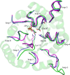
The comparison of the structures of the closed, 2PG bound complexes of wild-type TIM (1N55) (green), ml1TIM (1ML1, molecule A) (cyan) and A-TIM (2VEL, molecule B) (purple). ml1TIM has a shortened loop-3, and loop-1, as well as the point mutation A100W in loop-4. In A-TIM, loop-8 has also been shortened. There are no structural changes at the stability end of the TIM barrel, while at the catalytic end of the TIM-barrel significant structural changes in the loops are observed, due to the mutations/deletions in these loops
Fig. 9
Fig. 9
The alignment of some TIM sequences. Included in this sequence alignment are the sequences of well-studied triosephosphate isomerases: chicken (Gallus gallus), yeast (Saccharomyces cerevisiae), leish (Leishmania mexicana), tryp (Trypanosoma brucei brucei), ecoli (Escherichia coli), and pfal (Plasmodium falciparum). The catalytic residues are highlighted by asterisks, a filled square marks residues which on mutation are known to cause human diseases, the inverted filled triangle identifies the tip of the dimer interface loop-3, and a filled triangle highlights the four NH-groups providing the hydrogen bond donors interacting with the phosphate moiety oxygen atoms
Fig. 10
Fig. 10
The comparison of the mode of interactions of the hydroxamate reaction intermediate analogue and the active site residues of three sugar phosphate isomerases: TIM (cyan; 1TPH), RPI-B (green; 2BES) and PGI (magenta; 1KOJ) (in stereo). The hydroxamate moieties of each of the three structures have been superimposed. In each active site, the catalytic base is a glutamate: Glu167 in TIM, Glu75 in RPI-B and Glu357 in PGI. For the shared hydrogen bonding with the O1 and O2 atoms of the substrate and enediolate intermediate in TIM NE2(His95) is important, whereas in RPI-B and PGI the corresponding hydrogen bonding partners are OG(Ser71), and a water molecule (Wat241), respectively. The oxyanion hole for the O2 atom of the enediolate is made by Lys13 and His95 in TIM, by main chain NH-groups of residues Gly70-Ser71, as well as by Wat2064 and OG(Ser71) in RPI-B, and by three water molecules (Wat423, Wat477, Wat241) in PGI
Fig. 11
Fig. 11
The classical reaction mechanism. In this mechanism, the proton transfer steps are carried out by the glutamate as well as by the histidine
Fig. 12
Fig. 12
The criss-cross reaction mechanism. In this mechanism, all proton transfer steps are carried out by the glutamate side chain carboxylate group
Fig. 13
Fig. 13
Visualisation of the anisotropic B-factors of the atoms in the active site of the leishmanial TIM structure complexed with PGH (2VXN) (in stereo). The 70% probability thermal ellipsoids have been drawn. The highest B-factors are observed for OE1(Glu167), OE2(Glu167) and O1(PGH)
Fig. 14
Fig. 14
The comparison of the active site geometry of four liganded high resolution TIM structures, suggesting how a sliding motion of the Glu167 side chain over the catalytic end of the substrate could facilitate the proton shuttling by the catalytic base. The structures concern the complexes with PGH (2VXN) (purple), 2PG (1N55) (green), DHAP (1NEY) (orange), and IPP (1IF2) (cyan). In the PGH complex, the side chain is near Cys126, Ala165, Leu232, and in the IPP complex it is nearest to Ile172
Fig. 15
Fig. 15
The importance of Pro168 for the concerted loop-6/loop-7 closure. The active site geometry of the unliganded (open) conformation (5TIM, subunit A) (green) and the liganded (closed) conformation (1N55) (purple) are compared. The ligand is 2PG. On ligand binding, O(Gly211) (loop-7) rotates 90° upwards, clashing with the Pro168 (loop-6) side chain. Consequently, the Glu167-Pro168 dipeptide also switches to the closed conformation, by which the Glu167 side chain adopts the competent, swung-in conformation. This swung-in conformation is possible, once O(Gly211) has moved upwards

Similar articles

Cited by

References

    1. Knowles JR, Albery WJ. Perfection in enzyme catalysis: the energetics of triosephosphate isomerase. Acc Chem Res. 1977;10:105–111. doi: 10.1021/ar50112a001. - DOI
    1. Knowles JR. Enzyme catalysis: not different, just better. Nature. 1991;350:121–124. doi: 10.1038/350121a0. - DOI - PubMed
    1. Harris TK. The mechanistic ventures of triosephosphate isomerase. IUBMB Life. 2008;60:195–198. doi: 10.1002/iub.43. - DOI - PubMed
    1. Cui Q, Karplus M. Catalysis and specificity in enzymes: a study of triosephosphate isomerase and comparison with methyl glyoxal synthase. Adv Protein Chem. 2003;66:315–372. doi: 10.1016/S0065-3233(03)66008-0. - DOI - PubMed
    1. Albery WJ, Knowles JR. Efficiency and evolution of enzyme catalysis. Angew Chem Int Ed Engl. 1977;16:285–293. doi: 10.1002/anie.197702851. - DOI - PubMed

Publication types

MeSH terms

Substances

LinkOut - more resources