

Tailoring the degradation kinetics of mesoporous silicon structures through PEGylation
- PMID: 20694990
- PMCID: PMC2920054
- DOI: 10.1002/jbm.a.32807
Tailoring the degradation kinetics of mesoporous silicon structures through PEGylation
Abstract
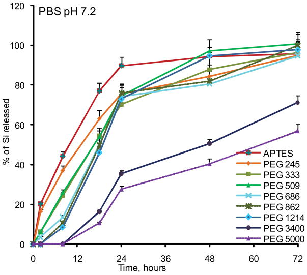
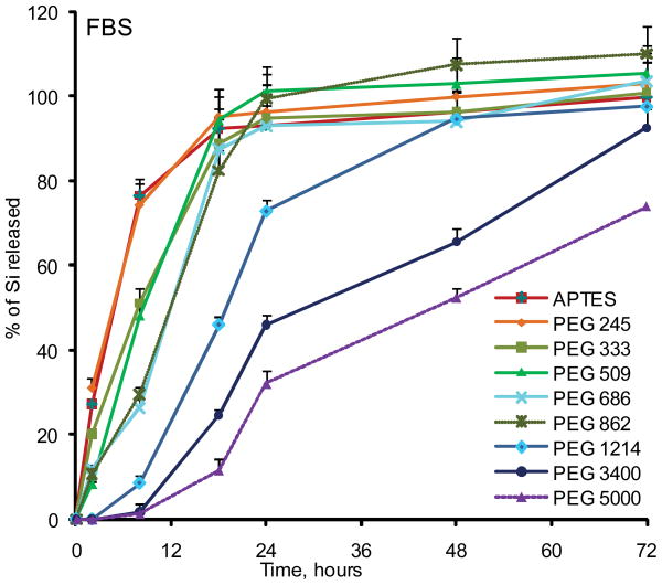
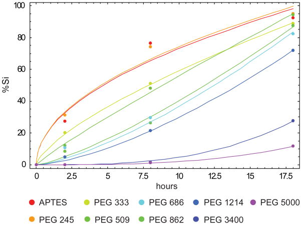
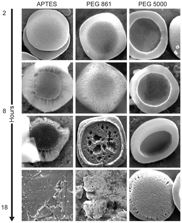
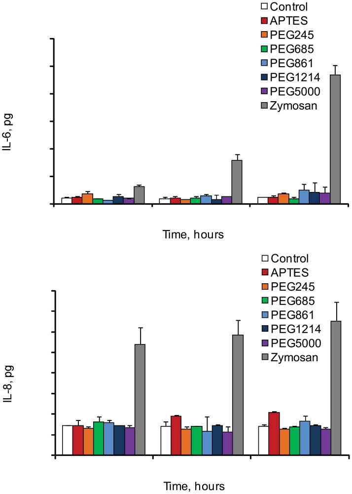
Injectable and implantable porosified silicon (pSi) carriers and devices for prolonged and controlled delivery of biotherapeutics offer great promise for treatment of various chronic ailments and acute conditions. Polyethylene glycols (PEGs) are important surface modifiers currently used in clinic mostly to avoid uptake of particulates by reticulo-endothelial system (RES). In this work we show for the first time that covalent attachment of PEGs to the pSi surface can be used as a means to tune degradation kinetics of silicon structures. Seven PEGs with varying molecular weights (245, 333, 509, 686, 1214, 3400, and 5000 Da) were employed and the degradation of PEGylated pSi hemispherical microparticles in simulated physiological conditions was monitored by means of ICP-AES, SEM, and fluorimetry. Biocompatibility of the systems with human macrophages in vitro was also evaluated. The results clearly indicate that controlled PEGylation of silicon microparticles can offer a sensitive tool to finely tune their degradation kinetics and that the systems do not induce release of proinflammatory cytokines IL-6 and IL-8 in THP1 human macrophages.
(c) 2010 Wiley Periodicals, Inc.
Figures

Similar articles
-
Functionalized mesoporous silicon for targeted-drug-delivery.Colloids Surf B Biointerfaces. 2012 Oct 1;98:18-25. doi: 10.1016/j.colsurfb.2012.04.018. Epub 2012 May 3. Colloids Surf B Biointerfaces. 2012. PMID: 22652355
-
Mitotic trafficking of silicon microparticles.Nanoscale. 2009 Nov;1(2):250-9. doi: 10.1039/b9nr00138g. Epub 2009 Oct 5. Nanoscale. 2009. PMID: 20644846 Free PMC article.
-
Silicon microfluidic flow focusing devices for the production of size-controlled PLGA based drug loaded microparticles.Int J Pharm. 2014 Jun 5;467(1-2):60-9. doi: 10.1016/j.ijpharm.2014.03.051. Epub 2014 Mar 28. Int J Pharm. 2014. PMID: 24680950
-
PEGylated bioactive molecules in biodegradable polymer microparticles.Expert Opin Biol Ther. 2007 Sep;7(9):1427-36. doi: 10.1517/14712598.7.9.1427. Expert Opin Biol Ther. 2007. PMID: 17727331 Review.
-
Multifunctional porous silicon for therapeutic drug delivery and imaging.Curr Drug Discov Technol. 2011 Sep;8(3):228-49. doi: 10.2174/157016311796799053. Curr Drug Discov Technol. 2011. PMID: 21291407 Review.
Cited by
-
Multistage vector (MSV) therapeutics.J Control Release. 2015 Dec 10;219:406-415. doi: 10.1016/j.jconrel.2015.08.010. Epub 2015 Aug 8. J Control Release. 2015. PMID: 26264836 Free PMC article. Review.
-
Bacteriophage Associated Silicon Particles: Design and Characterization of a Novel Theranostic Vector with Improved Payload Carrying Potential.J Mater Chem B. 2013 Oct 21;1(39):10.1039/C3TB20595A. doi: 10.1039/C3TB20595A. J Mater Chem B. 2013. PMID: 24409342 Free PMC article.
-
4D Multimodal Nanomedicines Made of Nonequilibrium Au-Fe Alloy Nanoparticles.ACS Nano. 2020 Oct 27;14(10):12840-12853. doi: 10.1021/acsnano.0c03614. Epub 2020 Sep 15. ACS Nano. 2020. PMID: 32877170 Free PMC article.
-
Site-Specific 111In-Radiolabeling of Dual-PEGylated Porous Silicon Nanoparticles and Their In Vivo Evaluation in Murine 4T1 Breast Cancer Model.Pharmaceutics. 2019 Dec 17;11(12):686. doi: 10.3390/pharmaceutics11120686. Pharmaceutics. 2019. PMID: 31861119 Free PMC article.
-
Mesoporous silicon (PSi) for sustained peptide delivery: effect of psi microparticle surface chemistry on peptide YY3-36 release.Pharm Res. 2012 Mar;29(3):837-46. doi: 10.1007/s11095-011-0611-6. Epub 2011 Oct 27. Pharm Res. 2012. PMID: 22033881
References
-
- Uhlir A. Electrolytic shaping of Germanium and Silicon. Bell System Technical Journal. 1956;35:333–347.
-
- Nijdam AJ, Ming-Cheng Cheng M, Geho DH, Fedele R, Herrmann P, Killian K, Espina V, Petricoin EF, 3rd, Liotta LA, Ferrari M. Physicochemically modified silicon as a substrate for protein microarrays. Biomaterials. 2007;28(3):550–8. - PubMed
-
- Lehmann V, Gosele U. Porous silicon formation: A quantum wire effect. Applied Physical Letters. 1991;58:656–658.
-
- Lin VS, Motesharei K, Dancil KP, Sailor MJ, Ghadiri MR. A porous silicon-based optical interferometric biosensor. Science. 1997;278(5339):840–3. - PubMed
Publication types
MeSH terms
Substances
Grants and funding
LinkOut - more resources
Full Text Sources
Other Literature Sources
Molecular Biology Databases
