Comparative Study
. 2010 Aug;124(3):233-51.
doi: 10.1037/a0019460.

A comparative view of face perception

Affiliations
Comparative Study

A comparative view of face perception

David A Leopold et al. J Comp Psychol. 2010 Aug.

Abstract

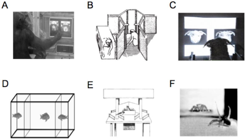
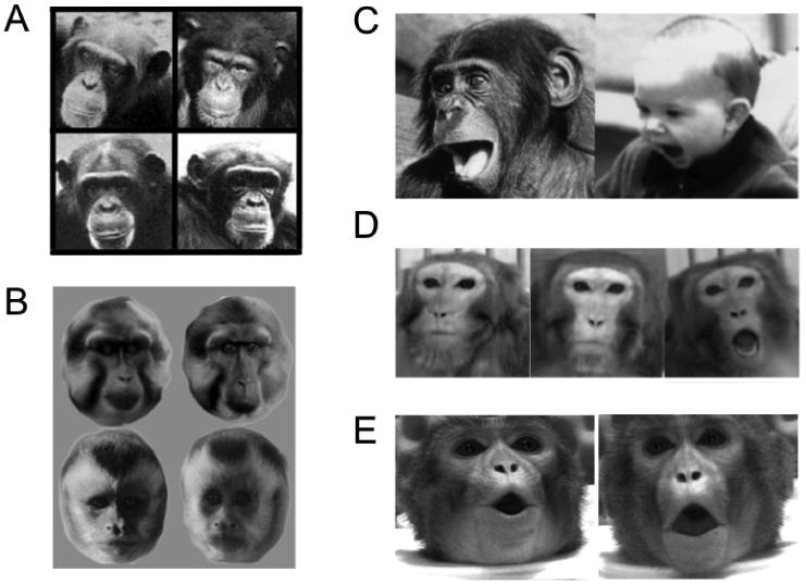
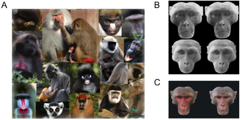
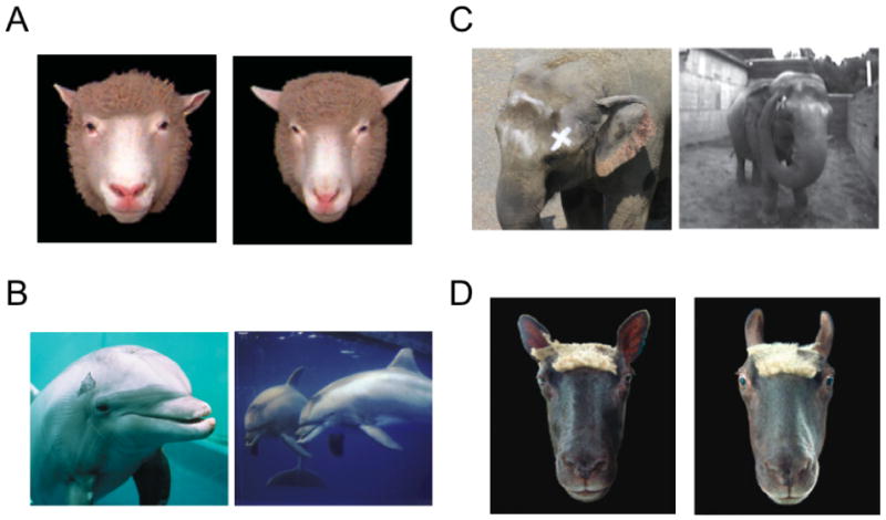
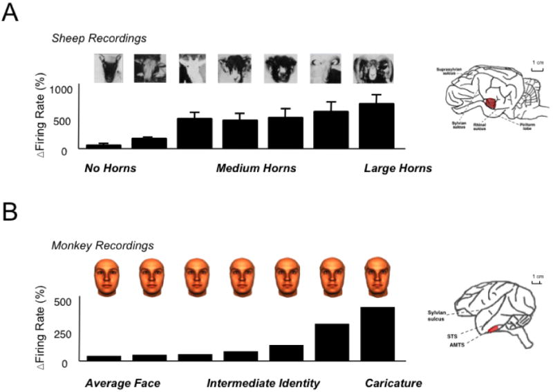
Face perception serves as the basis for much of human social exchange. Diverse information can be extracted about an individual from a single glance at their face, including their identity, emotional state, and direction of attention. Neuropsychological and functional magnetic resonance imaging (fMRI) experiments reveal a complex network of specialized areas in the human brain supporting these face-reading skills. Here we consider the evolutionary roots of human face perception by exploring the manner in which different animal species view and respond to faces. We focus on behavioral experiments collected from both primates and nonprimates, assessing the types of information that animals are able to extract from the faces of their conspecifics, human experimenters, and natural predators. These experiments reveal that faces are an important category of visual stimuli for animals in all major vertebrate taxa, possibly reflecting the early emergence of neural specialization for faces in vertebrate evolution. At the same time, some aspects of facial perception are only evident in primates and a few other social mammals, and may therefore have evolved to suit the needs of complex social communication. Because the human brain likely utilizes both primitive and recently evolved neural specializations for the processing of faces, comparative studies may hold the key to understanding how these parallel circuits emerged during human evolution.


Figures

Figure 1
Figure 1
Methods for testing animals on visual conspecific perception. A. A chimp indicates its recognition of a face by pressing on a touch-screen [1]. B. Macaque monkeys tested for capacity to visually recognize kin with whom they had no prior experience [2]. C. A sheep discriminates between two faces by pressing one of two panels in exchange for a food reward [3]. D. A fish inspects two neighbors in order to test whether it can subsequently recognize them [4]. E. Reactions of individual great tits to a radio-controlled maneuverable dummy was used to investigate the contribution of breast-stripe width to establishing social dominance [5]. F. A male jumping spider courts the video image of a female [6]. Figure 1 Citations [1] adapted from Martinez and Matsuzawa (2009). Animal Cognition, Suppl 1:S71-75. [2] adapted from Wu, H. M., Holmes, et al. (1980). Nature, 285(5762), 225-227. [3] adapted from Kendrick, K.M. (2008), in The Welfare of Sheep, (C. Dwyer, Ed.), Springer, Netherlands. [4] adapted from Balshine-Earn and Lotem (1997) Behaviour 135:369-386. [5] adapted from Jarvi and Bakken (1984) Animal Behaviour 32: 590-596. [6] adapted from Clark and Uetz (1990) Animal Behaviour. 40:884-890.
Figure 2
Figure 2
The recognition and interpretation of conspecific primate faces. A. Chimps can recognize familial face similarity between mothers (left column) and their sons (right column) [1]. B. Tonkean macaques and brown-faced capuchins are better able to discriminate between members of their own species (top and bottom rows, respectively) than between members of other species [2]. C. Homologous play face in bonobo and human [3]. D. Lip-smacking, neutral, and threat expressions from a rhesus macaque, used as stimuli for neurophysiological experiments [4]. E. Images taken from video stimuli shown to macaques with either matching or nonmatching acoustic vocalizations [5]. Figure 2 Citations [1] adapted from Parr and de Waal (1999) Nature, 399: 647-8. [2] adapted from DuFour et al (2006) Behavioral Processes, 73: 107-13. [3] adapted from Schmidt and Cohn (2001) Yearbook of Physical Anthropology, 44:8-24. [4] adapted from Gothard et al (2007) J Neurophysiology, 97:1671-1683. [5] adapted from Ghazanfar and Logothetis (2003) Nature, 423:937-8.
Figure 3
Figure 3
Face attributes associated with sexual selection in primates. A. Primate faces are often high contrast or colored, often with highlights around the eyes [1]. B. Both male and females rhesus monkeys prefer symmetrical (left column) over asymmetrical (right column) faces [2]. C. Male facial coloration in rhesus macaques affects viewing preference of females, with artificially reddened faces attracting longer periods of inspection [3]. Figure 3 Citations [1] adapted from Bradley B and Mundy N (2008). Evolutionary Anthropology, 17(2), 97-111. [2] adapted from Waitt C and Little AC (2006) International Journal of Primatology, 27(1),133-45. [3] adapted from Waitt C et al (2003) Proceedings of the Royal Society B: Biological Sciences, 270, S144-6.
Figure 4
Figure 4
The recognition of identity, expression, and self in mammals other than primates. A. Sheep (superorder Laurasiatheria) use the variation in the structure of the face to recognize one another [1]. B. Bottlenose dolphins (superorder Laurasiatheria) pass tests of self-recognition. When marked on the skin (left), they position themselves in front of the mirror in order to see the mark on their body (right) [2]. C. Similarly, elephants (superorder Afrotheria) pass tests of self-recognition. After receiving a mark on their face (left), they use their trunk in front of a mirror in an attempt to remove it (right) [3]. D. Sheep communicate emotion with their face, in this case indicating fear by drawing back the ears and opening the eyes [1]. Figure 4 Citations [1] adapted from Tate AJ et al (2006) Philosophical Transactions of the Royal Society B: Biological Sciences, 361: 2155-2172. [2] adapted from Marino et al (2007) PLOS Biology 5(5):966-972 and Reiss and Marino (2001) Proceedings of the National Academy of Sciences of the United States of America, 98:5937-42. [3] adapted from Plotnick et al (2006) Proceedings of the National Academy of Sciences of the United States of America, 103(45):17053-57.
Figure 5
Figure 5
Face processing in sheep and monkeys. (a) A subset of neurons in the sheep temporal cortex respond as a function of the visible length of the horns, a distinguishing feature for recognition of individual and species, as well as for social dominance [1]. (b) Neurons in the monkey temporal cortex respond to images of human faces, in this case responding monotonically to increasing amounts of “identity” level, or individuating feature information [2]. Figure 5 Citations. [1] adapted from Kendrick, KM (1994) Behavioral Processes 33:89-112. [2] Leopold DA et al (2006) Nature 442:572-575.
Figure 6
Figure 6
Visual analysis of conspecific faces in birds. (a) Budgerigars can discriminate pairs of real and synthetic conspecific faces. They are quicker at discriminating pairs of individuals when faces are configurally intact (left) than when they are scrambled (right) [1]. (b) The appearance of a nestling's open mouth determines the parental feeding response. Visual features include the size of the gape, internal patterning, and coloration [2]. Figure 6 Citations [1] adapted from Brown SD and Dooling RJ (1993) Journal of Comparative Psychology. 107(1):48-60. [2] adapted from Kilner RM et al (1999) Nature, 397:667-72.
Figure 7
Figure 7
Examples of invertebrate species with eyespot markings. (a) Owl butterfly with a conspicuous eyespot on its wing [1]. (b) Eyespot displays on the dorsum of a cuttlefish appear according to what type of predator is approaching [2]. (c) Caterpillar of the Great Orange Tip butterfly has false eyespots and the facial appearance of a green vine snake [3]. [1] adapted from http://en.wikipedia.org/wiki/Caligo_memnon. [2] adapted from Langridge KV et al (2007) Current Biology 17:R1044-R1045 [3] http://en.wikipedia.org/wiki/Hebomoia_glaucippe

Similar articles

Cited by

References

    1. Adachi I, Fujita K. Cross-modal representation of human caretakers in squirrel monkeys. Behavioural Processes. 2007;74(1):27–32. - PubMed
    1. Adolphs R. Neural systems for recognizing emotion. Current Opinions in Neurobiology. 2002;12(2):169–177. - PubMed
    1. Adolphs R, Gosselin F, Buchanan TW, Tranel D, Schyns P, Damasio AR. A mechanism for impaired fear recognition after amygdala damage. Nature. 2005;433(7021):68–72. - PubMed
    1. Adolphs R, Tranel D, Damasio AR. The human amygdala in social judgment. Nature. 1998;393(6684):470–474. - PubMed
    1. Allison T, Puce A, McCarthy G. Social perception from visual cues: role of the STS region. Trends in Cognitive Sciences. 2000;4(7):267–278. - PubMed

Publication types