

Investigation of the regenerative capacity of an acellular porcine medial meniscus for tissue engineering applications
- PMID: 20695759
- PMCID: PMC3011925
- DOI: 10.1089/ten.TEA.2009.0807
Investigation of the regenerative capacity of an acellular porcine medial meniscus for tissue engineering applications
Abstract
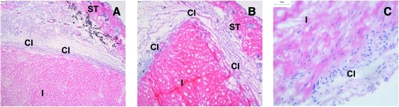
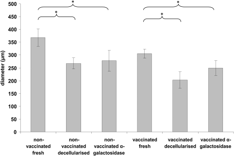
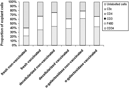
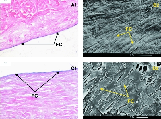
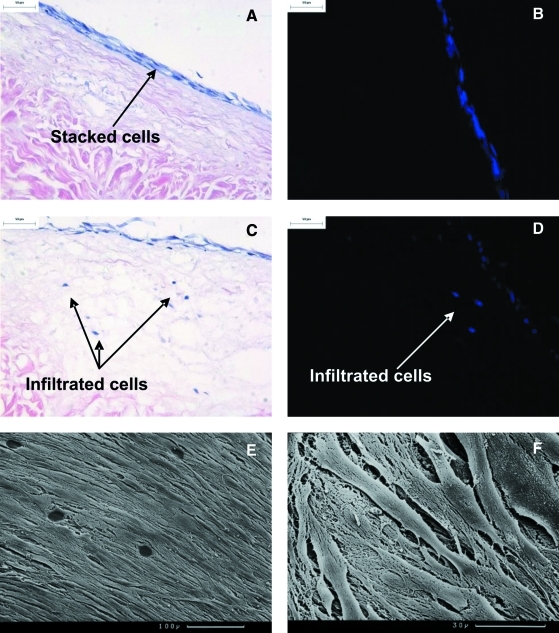
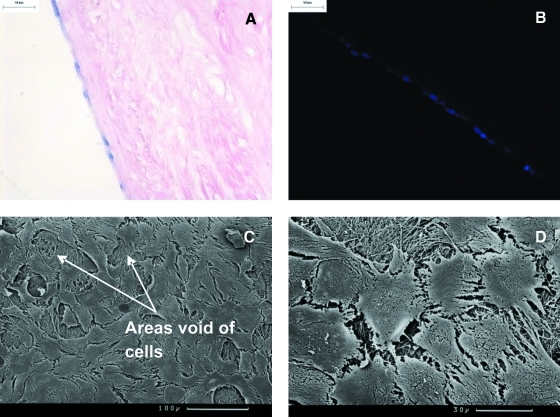
Previously, we have described the development of an acellular porcine meniscal scaffold. The aims of this study were to determine the immunocompatibility of the scaffold and capacity for cellular attachment and infiltration to gain insight into its potential for meniscal repair and replacement. Porcine menisci were decellularized by exposing the tissue to freeze-thaw cycles, incubation in hypotonic tris buffer, 0.1% (w/v) sodium dodecyl sulfate in hypotonic buffer plus protease inhibitors, nucleases, hypertonic buffer followed by disinfection using 0.1% (v/v) peracetic, and final washing in phosphate-buffered saline. In vivo immunocompatibility was assessed after implantation of the acellular meniscal scaffold subcutaneously into galactosyltransferase knockout mice for 3 months in comparison to fresh and acellular tissue treated with α-galactosidase (negative control). The cellular infiltrates in the explants were assessed by histology and characterized using monoclonal antibodies against: CD3, CD4, CD34, F4/80, and C3c. Static culture was used to assess the potential of acellular porcine meniscal scaffold to support the attachment and infiltration of primary human dermal fibroblasts and primary porcine meniscal cells in vitro. The explants were surrounded by capsules that were more pronounced for the fresh meniscal tissue compared to the acellular tissues. Cellular infiltrates compromised mononuclear phagocytes, CD34-positive cells, and nonlabeled fibroblastic cells. T-lymphocytes were sparse in all explanted tissue types and there was no evidence of C3c deposition. The analysis revealed an absence of a specific immune response to all of the implanted tissues. Acellular porcine meniscus was shown to be capable of supporting the attachment and infiltration of primary human fibroblasts and primary porcine meniscal cells. In conclusion, acellular porcine meniscal tissue exhibits excellent immunocompatibility and potential for cellular regeneration in the longer term.
Figures
Similar articles
-
Development and characterization of an acellular porcine medial meniscus for use in tissue engineering.Tissue Eng Part A. 2008 Apr;14(4):505-18. doi: 10.1089/tea.2007.0233. Tissue Eng Part A. 2008. PMID: 18370607
-
Tissue engineering small-diameter vascular grafts: preparation of a biocompatible porcine ureteric scaffold.Tissue Eng Part A. 2008 Nov;14(11):1871-82. doi: 10.1089/ten.tea.2007.0103. Tissue Eng Part A. 2008. PMID: 18950273
-
Biocompatibility and potential of acellular human amniotic membrane to support the attachment and proliferation of allogeneic cells.Tissue Eng Part A. 2008 Apr;14(4):463-72. doi: 10.1089/tea.2007.0145. Tissue Eng Part A. 2008. PMID: 18370928
-
Interactions of meniscal cells with extracellular matrix molecules: towards the generation of tissue engineered menisci.Cell Adh Migr. 2011 May-Jun;5(3):220-6. doi: 10.4161/cam.5.3.14463. Epub 2011 May 1. Cell Adh Migr. 2011. PMID: 21187716 Free PMC article. Review.
-
Animal models for meniscus repair and regeneration.J Tissue Eng Regen Med. 2015 May;9(5):512-27. doi: 10.1002/term.1760. Epub 2013 May 27. J Tissue Eng Regen Med. 2015. PMID: 23712959 Review.
Cited by
-
Antigen removal for the production of biomechanically functional, xenogeneic tissue grafts.J Biomech. 2014 Jun 27;47(9):1987-96. doi: 10.1016/j.jbiomech.2013.10.041. Epub 2013 Nov 8. J Biomech. 2014. PMID: 24268315 Free PMC article. Review.
-
Decellularized tissue and cell-derived extracellular matrices as scaffolds for orthopaedic tissue engineering.Biotechnol Adv. 2014 Mar-Apr;32(2):462-84. doi: 10.1016/j.biotechadv.2013.12.012. Epub 2014 Jan 10. Biotechnol Adv. 2014. PMID: 24417915 Free PMC article. Review.
-
A novel surgical technique for a rat subcutaneous implantation of a tissue engineered scaffold.Acta Histochem. 2018 Apr;120(3):282-291. doi: 10.1016/j.acthis.2018.02.010. Epub 2018 Mar 5. Acta Histochem. 2018. PMID: 29519681 Free PMC article.
-
Efficient decellularization for tissue engineering of the tendon-bone interface with preservation of biomechanics.PLoS One. 2017 Feb 7;12(2):e0171577. doi: 10.1371/journal.pone.0171577. eCollection 2017. PLoS One. 2017. PMID: 28170430 Free PMC article.
-
Stem cell delivery in tissue-specific hydrogel enabled meniscal repair in an orthotopic rat model.Biomaterials. 2017 Jul;132:59-71. doi: 10.1016/j.biomaterials.2017.04.004. Epub 2017 Apr 4. Biomaterials. 2017. PMID: 28407495 Free PMC article.
References
-
- Boyd K.T. Myers P.T. Meniscus preservation; rationale, repair techniques and results. Knee. 2003;10:1. - PubMed
-
- Baratz M.E. Fu F.H. Mengato R. Meniscal tears: the effect of meniscectomy and of repair on intraarticular contact areas and stress in the human knee. A preliminary report. Am J Sports Med. 1986;14:270. - PubMed
-
- Burke D.L. Ahmed A.H. Miller J. A biomechanical study of partial and total medial meniscectomy of the knee. Trans Orthop Res Soc. 1978;3:91.
-
- Altman G.H. Horan R.L. Lu H.H. Moreau J. Martin I. Richmond J.C. Kaplan D.L. Silk matrix for tissue engineered anterior cruciate ligaments. Biomaterials. 2002;23:4131. - PubMed
Publication types
MeSH terms
Substances
Grants and funding
LinkOut - more resources
Full Text Sources
Research Materials