

doi: 10.1111/j.1538-7836.2010.04006.x.
CLEC-2 is not required for platelet aggregation at arteriolar shear
Affiliations
- PMID: 20695981
- PMCID: PMC4362701
- DOI: 10.1111/j.1538-7836.2010.04006.x
Item in Clipboard
CLEC-2 is not required for platelet aggregation at arteriolar shear
J Thromb Haemost.
2010 Oct.
No abstract available
Figures
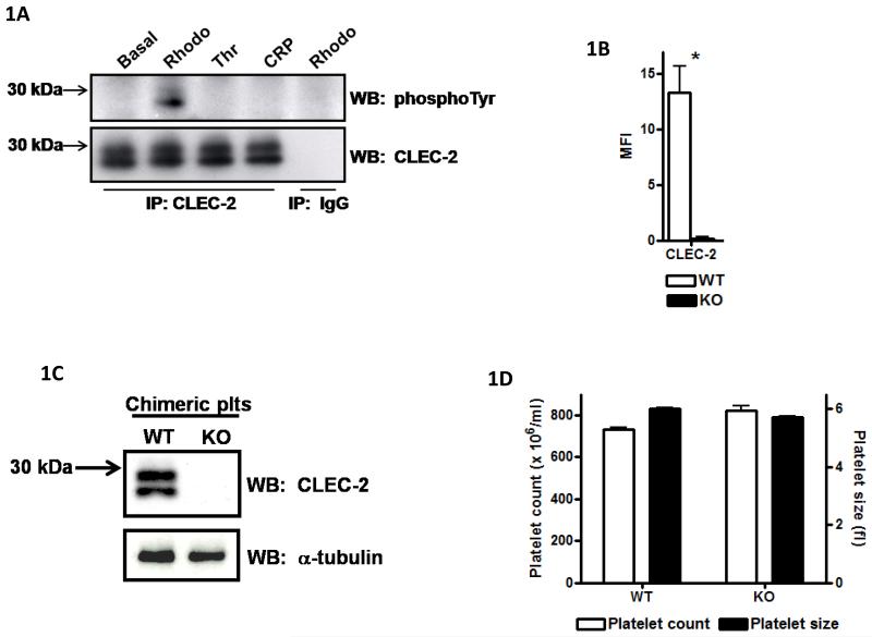
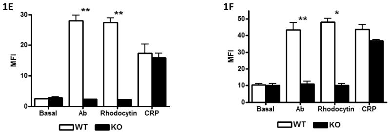
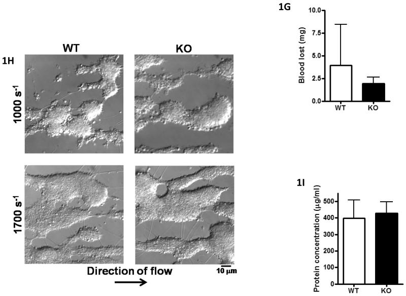
A) Washed platelets (5×10⁸/ml) under basal, rhodocytin (300nM), thrombin (1U/ml) or CRP-aggregated (3μg/ml) conditions were lysed with 2xNP40 lysis buffer. Lysates were immunoprecipitated with α-CLEC-2 or non-specific IgG antibodies and protein G sepharose. Precipitated proteins were separated by non-reducing SDS-PAGE and western blotted for phosphotyrosine and CLEC-2. Results are representative of 3 experiments. B) Washed platelets (2×10⁷/ml) were stained with 1μg α-CLEC-2 or non-specific IgG for 20min followed by staining with FITC-conjugated α-rat IgG for 20min in the dark. Platelets were analysed by flow cytometry and non-specific IgG readings subtracted. Results are from 8 experiments. *p<0.05. C) Washed platelets (2×10⁸/ml) were lysed with an equal volume of 2x non-reducing SDS sample buffer, separated by SDS-PAGE and western blotted for CLEC-2 and α-tubulin as a loading control. Results are representative of 3 experiments. D) Platelet number (×106/ml) and platelet size (fl) were analysed using a Blood Function Analyser. Results are from 8 experiments. E+F) Washed platelets (2×10⁷/ml) were stimulated with rhodocytin (100nM), α-CLEC-2 (10μg) or CRP (10μg/ml) for 2 min followed by staining with 5μl FITC-conjugated α-P-selectin antibody (E) or FITC-fibrinogen (F), for 30 min in the dark. Platelets were then analysed by flow cytometry. Results are from 3-4 experiments. **p<0.001, *p<0.05. G) A 0.2cm portion of tail was removed from anaesthetised mice. The assay was halted if blood loss had stopped for more than 5min. The amount of blood lost was determined by weight (shown ± standard deviation). Results are from 14 mice per group. H, I) Anticoagulated (to prevent thrombin generation) whole blood was passed through collagen-coated glass capillaries at a shear rate of 1000s−1 or 1700s−1 for 4 minutes. (H) Platelets were imaged by DIC microscopy then (I) lysed with NP40 lysis buffer and protein concentration determined (result shown for 1000s−1 only). Results are representative of 3-4 experiments.
A) Washed platelets (5×10⁸/ml) under basal, rhodocytin (300nM), thrombin (1U/ml) or CRP-aggregated (3μg/ml) conditions were lysed with 2xNP40 lysis buffer. Lysates were immunoprecipitated with α-CLEC-2 or non-specific IgG antibodies and protein G sepharose. Precipitated proteins were separated by non-reducing SDS-PAGE and western blotted for phosphotyrosine and CLEC-2. Results are representative of 3 experiments. B) Washed platelets (2×10⁷/ml) were stained with 1μg α-CLEC-2 or non-specific IgG for 20min followed by staining with FITC-conjugated α-rat IgG for 20min in the dark. Platelets were analysed by flow cytometry and non-specific IgG readings subtracted. Results are from 8 experiments. *p<0.05. C) Washed platelets (2×10⁸/ml) were lysed with an equal volume of 2x non-reducing SDS sample buffer, separated by SDS-PAGE and western blotted for CLEC-2 and α-tubulin as a loading control. Results are representative of 3 experiments. D) Platelet number (×106/ml) and platelet size (fl) were analysed using a Blood Function Analyser. Results are from 8 experiments. E+F) Washed platelets (2×10⁷/ml) were stimulated with rhodocytin (100nM), α-CLEC-2 (10μg) or CRP (10μg/ml) for 2 min followed by staining with 5μl FITC-conjugated α-P-selectin antibody (E) or FITC-fibrinogen (F), for 30 min in the dark. Platelets were then analysed by flow cytometry. Results are from 3-4 experiments. **p<0.001, *p<0.05. G) A 0.2cm portion of tail was removed from anaesthetised mice. The assay was halted if blood loss had stopped for more than 5min. The amount of blood lost was determined by weight (shown ± standard deviation). Results are from 14 mice per group. H, I) Anticoagulated (to prevent thrombin generation) whole blood was passed through collagen-coated glass capillaries at a shear rate of 1000s−1 or 1700s−1 for 4 minutes. (H) Platelets were imaged by DIC microscopy then (I) lysed with NP40 lysis buffer and protein concentration determined (result shown for 1000s−1 only). Results are representative of 3-4 experiments.
A) Washed platelets (5×10⁸/ml) under basal, rhodocytin (300nM), thrombin (1U/ml) or CRP-aggregated (3μg/ml) conditions were lysed with 2xNP40 lysis buffer. Lysates were immunoprecipitated with α-CLEC-2 or non-specific IgG antibodies and protein G sepharose. Precipitated proteins were separated by non-reducing SDS-PAGE and western blotted for phosphotyrosine and CLEC-2. Results are representative of 3 experiments. B) Washed platelets (2×10⁷/ml) were stained with 1μg α-CLEC-2 or non-specific IgG for 20min followed by staining with FITC-conjugated α-rat IgG for 20min in the dark. Platelets were analysed by flow cytometry and non-specific IgG readings subtracted. Results are from 8 experiments. *p<0.05. C) Washed platelets (2×10⁸/ml) were lysed with an equal volume of 2x non-reducing SDS sample buffer, separated by SDS-PAGE and western blotted for CLEC-2 and α-tubulin as a loading control. Results are representative of 3 experiments. D) Platelet number (×106/ml) and platelet size (fl) were analysed using a Blood Function Analyser. Results are from 8 experiments. E+F) Washed platelets (2×10⁷/ml) were stimulated with rhodocytin (100nM), α-CLEC-2 (10μg) or CRP (10μg/ml) for 2 min followed by staining with 5μl FITC-conjugated α-P-selectin antibody (E) or FITC-fibrinogen (F), for 30 min in the dark. Platelets were then analysed by flow cytometry. Results are from 3-4 experiments. **p<0.001, *p<0.05. G) A 0.2cm portion of tail was removed from anaesthetised mice. The assay was halted if blood loss had stopped for more than 5min. The amount of blood lost was determined by weight (shown ± standard deviation). Results are from 14 mice per group. H, I) Anticoagulated (to prevent thrombin generation) whole blood was passed through collagen-coated glass capillaries at a shear rate of 1000s−1 or 1700s−1 for 4 minutes. (H) Platelets were imaged by DIC microscopy then (I) lysed with NP40 lysis buffer and protein concentration determined (result shown for 1000s−1 only). Results are representative of 3-4 experiments.
References
-
- May F, Hagedorn I, Pleines I, Bender M, Vogtle T, Eble J, Elvers M, Nieswandt B. CLEC-2 is an essential platelet-activating receptor in hemostasis and thrombosis. Blood. 2009;114:3464–72. - PubMed
-
- Suzuki-Inoue K, Fuller GL, Garcia A, Eble JA, Pohlmann S, Inoue O, Gartner TK, Hughan SC, Pearce AC, Laing GD, Theakston RD, Schweighoffer E, Zitzmann N, Morita T, Tybulewicz VL, Ozaki Y, Watson SP. A novel Syk-dependent mechanism of platelet activation by the C-type lectin receptor CLEC-2. Blood. 2006;107:542–9. - PubMed
-
- Suzuki-Inoue K, Kato Y, Inoue O, Kaneko MK, Mishima K, Yatomi Y, Yamazaki Y, Narimatsu H, Ozaki Y. Involvement of the snake toxin receptor CLEC-2, in podoplanin-mediated platelet activation, by cancer cells. J Biol Chem. 2007;282:25993–6001. - PubMed
Publication types
MeSH terms
Substances
Grants and funding
LinkOut - more resources
Full Text Sources
Molecular Biology Databases
