

Comparison of laser Doppler imaging, fingertip lacticemy test, and nailfold capillaroscopy for assessment of digital microcirculation in systemic sclerosis
- PMID: 20696074
- PMCID: PMC2945058
- DOI: 10.1186/ar3112
Comparison of laser Doppler imaging, fingertip lacticemy test, and nailfold capillaroscopy for assessment of digital microcirculation in systemic sclerosis
Abstract
Introduction: Laser Doppler imaging (LDI) is a relatively new method for assessing the functional aspect of superficial skin blood flow in systemic sclerosis (SSc) and Raynaud's phenomenon. The present study investigated the dynamic behavior of digital skin microvascular blood flow before and after cold stimulus (CS) in SSc patients and in healthy controls by means of a comprehensive approach of the functional (LDI), morphological (nailfold capillaroscopy (NFC)), and biochemical (fingertip lacticemy (FTL)) microcirculation components.
Methods: Forty-four SSc patients and 40 healthy controls were included. After acclimatization, all subjects underwent NFC followed by LDI and FTL measurement. NFC was performed with a stereomicroscope under 10× to 20× magnification in the 10 digits of the hands. Skin blood flow of the dorsum of four fingertips (excluding the thumb) of the left hand was measured using LDI at baseline and for 30 minutes after CS. The mean finger blood flow (FBF) of the four fingertips was expressed as arbitrary perfusion units. FTL was determined on the fourth left finger before (pre-CS-FTL) and 10 minutes after CS.
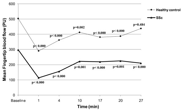
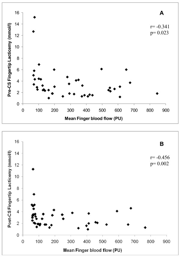
Results: LDI showed significantly lower mean baseline FBF in SSc patients as compared with controls (296.9 ± 208.8 vs. 503.6 ± 146.4 perfusion units; P < 0.001) and also at all time points after CS (P < 0.001). There was a significant decrease in mean FBF after CS as compared with baseline in SSc patients and in controls, followed by recovery of the blood flow 27 minutes after CS in healthy controls, but not in SSc patients. FBF tended to be lower in patients with digital scars and previous ulceration/amputation (P = 0.06). There was no correlation between mean baseline FBF and NFC parameters. Interestingly, there was a negative correlation between FTL and FBF measured by LDI in basal conditions and 10 minutes after CS in SSc patients.
Conclusions: LDI showed lower digital blood flow in SSc patients when compared with healthy controls and correlated well with FTL both at baseline and after CS, allowing objective measurement of blood perfusion in SSc patients. The lack of correlation between functional and morphological microvascular abnormalities, measured by LDI and NFC, suggests they are complementary tools for evaluation of independent microangiopathy aspects in SSc patients.
Figures
Comment in
-
Connective tissue diseases: Blood flow assessment at your fingertips.Nat Rev Rheumatol. 2010 Oct;6(10):555. doi: 10.1038/nrrheum.2010.146. Nat Rev Rheumatol. 2010. PMID: 20925149 No abstract available.
-
Response to: comparison of laser Doppler imaging, fingertip lacticemy test, and nailfold capillaroscopy for assessment of digital microcirculation in systemic sclerosis.Arthritis Res Ther. 2011 Jan 24;13(1):301; author reply 302. doi: 10.1186/ar3201. Arthritis Res Ther. 2011. PMID: 21345273 Free PMC article. No abstract available.
References
-
- Maricq HR, Weinrich MC, Valter I, Palesch YY, Maricq JG. Digital vascular responses to cooling in subjects with cold sensitivity, primary Raynaud's phenomenon, or scleroderma spectrum disorders. J Rheumatol. 1996;23:2068–2078. - PubMed
-
- Maricq HR, McGregor AR, Diat F, Smith EA, Maxwell DB, LeRoy EC, Weinrich MC. Major clinical diagnoses found among patients with raynaud Phenomenon from the General Population. J Rheumatol. 1990;17:1171–1176. - PubMed
Publication types
MeSH terms
LinkOut - more resources
Full Text Sources
Medical
Miscellaneous
