. 2010 Aug 10:10:415.
doi: 10.1186/1471-2407-10-415.

TLR9 expression in glioma tissues correlated to glioma progression and the prognosis of GBM patients

Affiliations

TLR9 expression in glioma tissues correlated to glioma progression and the prognosis of GBM patients

Chao Wang et al. BMC Cancer. .

Abstract

Background: Our study aims to evaluate the expression of TLR9 in glioma tissues, examine the association between TLR9 expression, clinicopathological variables, and glioma patient outcome, we further characterized the direct effects of TLR9 agonist CpG ODN upon the proliferation and invasion of glioma cells in vitro.

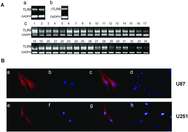
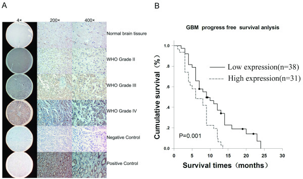
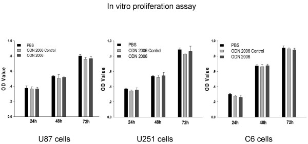
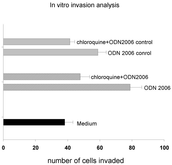
Methods: RT-PCR and immunofluorescence were used to determine the expression of TLR9 in glioma cell lines and clinical glioma samples. Tissue microarry and immunohistochemistry were applied to evaluated TLR9 expression in 292 newly diagnosed glioma and 13 non-neoplastic brain tissues. We further investigated the effect of CpG ODN on the proliferation and invasion of glioma cells in vitro with MTT assays and matrigel transwell assay respectively.

Results: RT-PCR showed that TLR9 expressed in all the glioma samples and glioma cell lines we examined. The tissue array analysis indicated that TLR9 expression is correlated with malignancy of glioma (p < 0.01). Multivariate Cox regression analysis revealed that TLR9 expression is an independent prognostic factor for PFS of GBM patients (P = 0.026). TLR9 agonist CpG ODN has no significant effect on glioma proliferation, but matrigel transwell analysis showed that TLR9 agonist CpG ODN can significantly enhance glioma invasion in vitro.

Conclusions: Our data indicated that TLR9 expression increases according to the histopathological grade of glioma, and the TLR9 expression level is related to the PFS of GBM patients. In addition, our findings warrant caution in the directly injection of TLR9 agonist CpG ODN into glioma tissues for the glioma immunotherapy.


Figures

Figure 1
Figure 1
Expression of TLR9 in glioma cell lines and glioma samples. A. mRNA expression of TLR9 (a) Expression of TLR9 in human U87 glioma cell line(lane1) and human U251 glioma cell line(lane2); (b) Expression of TLR9 in rat glioma cell line C6. (c) Expression of TLR9 in 34 clinical glioma samples (lane1-15, Grade IV gliomas; lane16-28, grade III gliomas;lane 29-34, grade II gliomas). GAPDH was used as positive control. B. Protein expression of TLR9 in human glioma cell lines U87 and U251 with immunofluorescence staining. cytoplasmic localization of TLR9(red fluorescence, Cy3) in U87 and U251 was confirmed by confocal microscopy, nucleus was stained with Hoechst(Blue fluorescence). (a) TLR9 staining in U87 cells. (b) Hoechst staining of nucleus in U87 cells. (c) The combined figure of a and b. (d) negative control, staining of U87 cells with secondary antibody and Hoechst, but without primary anti-TLR9 antibody. (e) TLR9 staining in U251 cells. (f) Hoechst staining of nucleus in U251 cells. (g) The combined figure of e and f. (h) Negative control, staining of U251 cells with secondary antibody and Hoechst, but without primary anti-TLR9 antibody.
Figure 2
Figure 2
TLR9 expression in glioma tissues correlated to glioma progression and the prognosis of GBM patients. A. Representative immunohistochemistry staining photos of the glioma tissue arrays. Normal human kidney tissues provided by Department of Surgery were used as positive control. For negative controls, the primary antibody was replaced by normal mouse serum. Cell nuclei were counterstained with haematoxylin. B. Kaplan-Meier analysis showing the PFS(progress free survival) of glioblatoma (GBM) patients in low TLR9 expression group (bold line) and high TLR9 expression group (dotted line). The difference of PFS between the two groups is significant(p = 0.001).
Figure 3
Figure 3
Proliferation of glioma cell lines in response to CpG ODN(ODN 2006). Human U87 glioma cell line, human U251 glioma cell line, and rat C6 glioma cell line were incubated with CpG ODN(10 uM), CpG ODN Control(10 uM) or same volume of PBS. The analysis was performed in triplicate and cell growth was measured at 24, 48, 72 hours with an ELISA reader at 490 nm, data was represented as OD value. Bars refer to the standard error of the mean value. Comparison between groups was made by using student's t test, P ≤ 0.05 was defined as significant.
Figure 4
Figure 4
Invasion of GBM cell line U87 in response to CpG ODN(ODN 2006). U87 cells were plated in 24 well cell culture chambers using transwell inserts precotated with matrigel. ODN 2006 (10 uM), ODN 2006 control (10 uM), same volume of the control medium, or TLR signaling inhibitor chloroquine (10 uM) were added and incubated for 24 hours, the number of invaded cells was stained and counted, all analysis were performed in triplicate. The invasion ability was represented as numbers of invaded cells. Bars refer to the standard error of the mean value. Comparison between groups was made by using student's t test, P ≤ 0.05 was defined as significant.

References

    1. Bondy ML, Scheurer ME, Malmer B, Barnholtz-Sloan JS, Davis FG, Il'yasova D, Kruchko C, McCarthy BJ, Rajaraman P, Schwartzbaum JA. Brain tumor epidemiology: consensus from the Brain Tumor Epidemiology Consortium. Cancer. 2008;113(7 Suppl):1953–1968. doi: 10.1002/cncr.23741. - DOI - PMC - PubMed
    1. Walker PR, Calzascia T, de Tribolet N, Dietrich PY. T-cell immune responses in the brain and their relevance for cerebral malignancies. Brain Res Brain Res Rev. 2003;42(2):97–122. doi: 10.1016/S0165-0173(03)00141-3. - DOI - PubMed
    1. Vollmer J, Krieg AM. Immunotherapeutic applications of CpG oligodeoxynucleotide TLR9 agonists. Adv Drug Deliv Rev. 2009;61(3):195–204. doi: 10.1016/j.addr.2008.12.008. - DOI - PubMed
    1. Kadowaki N, Ho S, Antonenko S, Malefyt RW, Kastelein RA, Bazan F, Liu YJ. Subsets of human dendritic cell precursors express different toll-like receptors and respond to different microbial antigens. J Exp Med. 2001;194(6):863–869. doi: 10.1084/jem.194.6.863. - DOI - PMC - PubMed
    1. Boonstra A, Rajsbaum R, Holman M, Marques R, Asselin-Paturel C, Pereira JP, Bates EEM, Akira S, Vieira P, Liu Y-J. Macrophages and Myeloid Dendritic Cells, but Not Plasmacytoid Dendritic Cells, Produce IL-10 in Response to MyD88- and TRIF-Dependent TLR Signals, and TLR-Independent Signals. J Immunol. 2006;177(11):7551–7558. - PubMed

Publication types

MeSH terms