

Review
doi: 10.1016/j.febslet.2010.08.006.
Epub 2010 Aug 7.
Mast cell signaling: the role of protein tyrosine kinase Syk, its activation and screening methods for new pathway participants
Affiliations
- PMID: 20696166
- PMCID: PMC2998559
- DOI: 10.1016/j.febslet.2010.08.006
Item in Clipboard
Review
Mast cell signaling: the role of protein tyrosine kinase Syk, its activation and screening methods for new pathway participants
FEBS Lett.
.
Abstract
The aggregation by antigen of the IgE bound to its high affinity receptor on mast cells initiates a complex series of biochemical events that result in the release of inflammatory mediators. The essential role of the protein tyrosine kinase Syk has been appreciated for some time, and newer results have defined the mechanism of its activation. The use of siRNA has defined the relative contribution of Syk, Fyn and Gab2 to signaling and has made possible a screening study to identify previously unrecognized molecules that are involved in these pathways.
Published by Elsevier B.V.
Figures
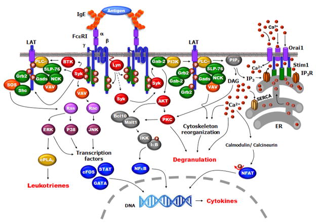
Model Depicting major molecules and events in mast cell activation. Antigen binding to IgE cross-links two FcεRI leading to Lyn phosphorylating the tyrosines in the ITAMs of the β and γ-subunits of the receptor. Syk kinase is recruited and activated by binding to the phosphorylated ITAM of the γ subunit; the activated Syk autophosphorylates and activates downstream signaling which includes phosphorylation of Btk, PI3K, the adaptors LAT1, LAT2, SLP-76 and PLCγ. Multimolecular complexes form on LAT which includes Gads, SLP76, PLCγ and Vav. The activated PLCγ hydrolyzes PI(4,5)P2 to form DAG and IP3. The IP3 activates the IP3-receptor on the ER releasing calcium, the calcium sensor STIM1 then interacts with the ORA1 membrane protein opening the CRAC channels allowing the increase in intracellular calcium. DAG activates PKC and also interacts with calmodulin to activate calcineurin leading to NFAT translocation into the nucleus. The activation of GTPases lead to MAP kinase activation. These pathways lead to degranulation, release of leukotrienes and cytokine synthesis.
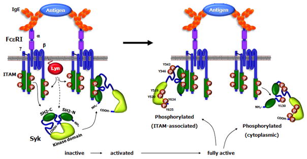
Syk Activation. The globular representation of Syk shows the molecule in its autoinhibitory state with the COOH-terminal tail interacting with the inter-SH2 domains thereby contributing to keeping the molecule in a closed conformation. The binding of Syk to the phosphorylated ITAM results in a conformational change that exposes the COOH-terminal region. This leads to the phosphorylation of two tyrosines in the tail, which then keeps the molecule in an open conformation allowing for further phosphorylation of other tyrosine residues mostly by autophosphorylation, although there could be contributions by other tyrosine kinases. Phosphorylation of Tyr-130 in the inter-SH2 domain results in Syk dissociating from the ITAM.
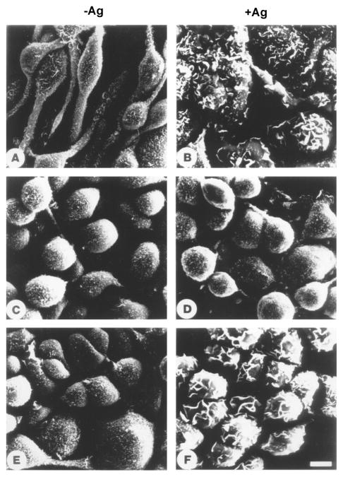
The FcεRI-induced morphological changes in mast cells require Syk. RBL-2H3 (Syk+), TB1A2 (Syk−), and the Syk transfected 3A5 cell line were cultured with antigen specific IgE, washed and then either incubated with medium alone (A,C,E) or activated with antigen (B,D,F). After 20 minutes the cells were prepared for scanning electron microscopy. The nonstimulated RBL-2H3 cells are spindle-shaped with their surface covered with small short microvilli (A); FcεRI activation induced cell spreading and transformed the cell surface to a lamellar topography with deep folds and ruffles (B). The Syk-negative variant are less spindle-shaped and more round and transfection of Syk into these cells did not change their appearance (C and E). FcεRI aggregation did not induce any morphological changes in the Syk-negative cells (D) while these receptor-induced morphological changes are reconstituted by transfection of Syk (F).
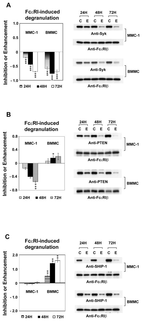
The comparisons of targeting Syk, PTEN and SHIP-1 in MMC-1 and BMMC. The mouse mast cells line (MMC-1), or Bone Marrow Derived Mast Cells (BMMC) were transfected with siRNA for Syk (A), PTEN (B) or SHIP-1 (C). Control cells were transfected with scrambled siRNA “C”. The cells were sensitized and then stimulated with antigen after 24, 48 and 72 hours. The antigen-induced β-hexosaminidase release is expressed as the fraction of that in controls. The lysates of the cell pellets were analyzed by immunoblotting with the indicated antibodies using the anti-FcεRIβ as a loading control. “C” for scrambled siRNA controls, “E” experimental siRNA.
References
-
- Siraganian RP. Mast cell signal transduction from the high-affinity IgE receptor. Curr Opin Immunol. 2003;15:639–46. - PubMed
-
- Zhang J, Billingsley ML, Kincaid RL, Siraganian RP. Phosphorylation of Syk activation loop tyrosines is essential for Syk function. An in vivo study using a specific anti-Syk activation loop phosphotyrosine antibody. J Biol Chem. 2000;275:35442–7. - PubMed
Publication types
MeSH terms
Substances
Grants and funding
LinkOut - more resources
Full Text Sources
Other Literature Sources
Miscellaneous
