

Hypercholesterolemia in rats impairs the cholinergic system and leads to memory deficits
- PMID: 20696249
- PMCID: PMC2977849
- DOI: 10.1016/j.mcn.2010.08.001
Hypercholesterolemia in rats impairs the cholinergic system and leads to memory deficits
Abstract
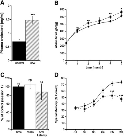
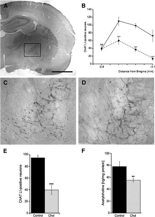
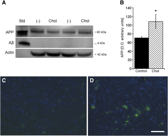
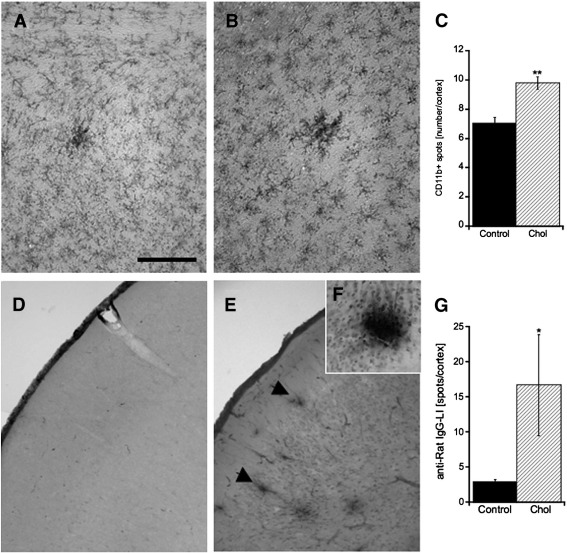
Alzheimer's disease (AD) is a chronic brain disorder characterized by cognitive impairment, cholinergic dysfunction, inflammation, tau and beta-amyloid pathology and vascular damage. Recent studies have shown, that high cholesterol levels are linked to the pathology of AD. The aim of our present work was to study the effects of hypercholesterolemia in adult rats. Five months after 5% cholesterol-enriched diet plasma cholesterol levels and total weight were significantly enhanced compared to controls. Spatial memory was studied in an 8-arm radial maze and cholesterol-treated rats showed an impaired learning and long-term memory. Hypercholesterolemia significantly reduced the number of cholinergic neurons in the basal nucleus of Meynert and decreased acetylcholine levels in the cortex. Nerve growth factor was only slightly enhanced in the cortex of cholesterol-treated animals. Levels of amyloid precursor protein, beta-amyloid(1-42), as well as tau and phospho-tau 181 were significantly enhanced in the cortex of cholesterol-fed rats. Hypercholesterolemia markedly increased several cerebral inflammatory markers and enhanced microglial CD11b-like immunoreactivity. Vascular density, stained by RECA-1 was not changed. However, cholesterol induced cortical microbleedings illustrated by intensive anti-rat IgG-positive spots in the cortex. In conclusion, our data demonstrate that hypercholesterolemia in rats caused memory impairment, cholinergic dysfunction, inflammation, enhanced cortical beta-amyloid and tau and microbleedings, all indications, which resemble an AD-like pathology.
Copyright © 2010 Elsevier Inc. All rights reserved.
Figures
Similar articles
-
Hypercholesterolemia accelerates intraneuronal accumulation of Aβ oligomers resulting in memory impairment in Alzheimer's disease model mice.Life Sci. 2012 Dec 10;91(23-24):1169-76. doi: 10.1016/j.lfs.2011.12.022. Epub 2012 Jan 17. Life Sci. 2012. PMID: 22273754
-
Hypercholesterolemia accelerates amyloid β-induced cognitive deficits.Int J Mol Med. 2013 Mar;31(3):577-82. doi: 10.3892/ijmm.2013.1233. Epub 2013 Jan 8. Int J Mol Med. 2013. PMID: 23314909
-
High cholesterol-induced neuroinflammation and amyloid precursor protein processing correlate with loss of working memory in mice.J Neurochem. 2008 Jul;106(1):475-85. doi: 10.1111/j.1471-4159.2008.05415.x. Epub 2008 Jul 1. J Neurochem. 2008. PMID: 18410513 Free PMC article.
-
The significance of the cholinergic system in the brain during aging and in Alzheimer's disease.J Neural Transm (Vienna). 2006 Nov;113(11):1625-44. doi: 10.1007/s00702-006-0579-2. Epub 2006 Oct 13. J Neural Transm (Vienna). 2006. PMID: 17039298 Review.
-
The cholinergic system in aging and neuronal degeneration.Behav Brain Res. 2011 Aug 10;221(2):555-63. doi: 10.1016/j.bbr.2010.11.058. Epub 2010 Dec 9. Behav Brain Res. 2011. PMID: 21145918 Review.
Cited by
-
The sterol regulatory element-binding protein 2 is dysregulated by tau alterations in Alzheimer disease.Brain Pathol. 2019 Jul;29(4):530-543. doi: 10.1111/bpa.12691. Epub 2019 Jan 28. Brain Pathol. 2019. PMID: 30515907 Free PMC article.
-
Metabolic Disturbance of High-Saturated Fatty Acid Diet in Cognitive Preservation.Int J Mol Sci. 2023 Apr 28;24(9):8042. doi: 10.3390/ijms24098042. Int J Mol Sci. 2023. PMID: 37175748 Free PMC article.
-
Lipidopathy disrupts peripheral and central amyloid clearance in Alzheimer's disease: Where are our knowledge.IBRO Neurosci Rep. 2025 Jan 9;18:191-199. doi: 10.1016/j.ibneur.2025.01.004. eCollection 2025 Jun. IBRO Neurosci Rep. 2025. PMID: 39906286 Free PMC article. Review.
-
Effects of oxidative stress on amyloid precursor protein processing in rat and human platelets.Platelets. 2013;24(1):26-36. doi: 10.3109/09537104.2012.661104. Epub 2012 Mar 2. Platelets. 2013. PMID: 22385218 Free PMC article.
-
High‑fat treatment prevents postoperative cognitive dysfunction in a hyperlipidemia model by protecting the blood‑brain barrier via Mfsd2a‑related signaling.Mol Med Rep. 2019 Nov;20(5):4226-4234. doi: 10.3892/mmr.2019.10675. Epub 2019 Sep 12. Mol Med Rep. 2019. PMID: 31545471 Free PMC article.
References
-
- Aihara N., Tanno H., Hall J.J., Pitts L.H., Noble L.J. Immunocytochemical loclization of immunoglobulins in the rat brain: relationship to the blood-brain barrier. J. Comp. Neurol. 1994;342:481–496. - PubMed
-
- Akiyama H., Barger S., Barnum S., Bradt B., Bauer J., Cole G.M., Cooper N.R., Eikelenboom P., Emmerling M., Fiebich B.L., Finch C.E., Frautschy S., Griffin W.S.T., Hampel H., Hull M., Landreth G., Lue L.-F., Mrak R., Mackenzie I.R., McGeer P.L., O'Bannion M.K., Pachter J., Pasinetti G., Plata-Salaman C., Rogers J., Rydel R., Shen Y., Streit W., Strohmeyer R., Tooyoma I., Van Muiswinkel F.L., Veerhuis R., Walker D., Webster S., Wegrzyniak B., Wenk G., Wyss-Coray T. Inflammation and Alzheimer's disease. Neurobiol. Aging. 2000;21:383–421. - PMC - PubMed
-
- Anstey K.J., Lipnicki D.M., Low L.F. Cholesterol as a risk factor for dementia and cognitive decline: a systematic review of prospective studies with meta-analysis. Am. J. Geriatr. Psychiatry. 2008;16:343–354. - PubMed
-
- Barnett J.V., Haigh L.S., Marsh J.D., Galper J.B. Effects of low density lipoproteins and mevinolin on sympathetic responsiveness in cultured chick atrial cells. J. Biol. Chem. 1989;264:10779–10786. - PubMed
Publication types
MeSH terms
Substances
Grants and funding
LinkOut - more resources
Full Text Sources
Medical
Research Materials
