Review
. 2010 Nov 28;703(1):18-23.
doi: 10.1016/j.mrgentox.2010.07.013. Epub 2010 Aug 7.

DNA polymerase structure-based insight on the mutagenic properties of 8-oxoguanine

Affiliations
Review

DNA polymerase structure-based insight on the mutagenic properties of 8-oxoguanine

William A Beard et al. Mutat Res. .

Abstract

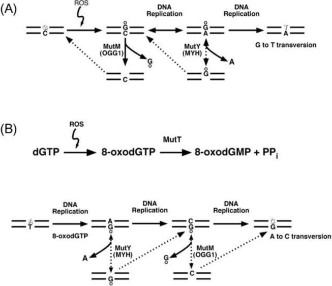
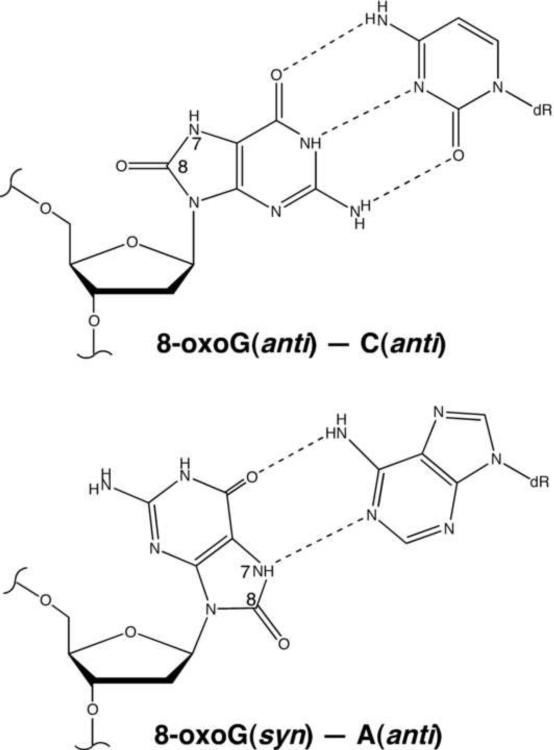
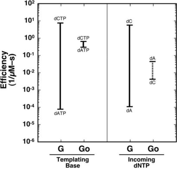
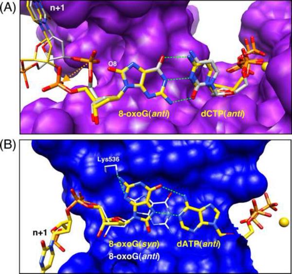
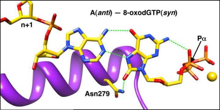
An aerobic environment burdens DNA polymerase substrates with oxidized substrates (DNA and nucleotide pools). A major promutagenic lesion resulting from oxidative stress is 8-oxo-7,8-dihydro-2'-deoxyguanosine (8-oxoG). Guanine oxidation alters the hydrogen bonding properties of the base and glycosidic-preference of the nucleotide. The favored glycosidic syn-conformation exposes the Hoogsteen edge of the base for hydrogen bonding with adenine during DNA synthesis. The cell has recognized the threat of this lesion and has evolved an intricate surveillance system to provide DNA polymerases with unmodified substrates. Failure to do so leads to transversion mutations. Since the mutagenic properties of the base are dictated by the anti-syn-conformation of the nucleotide, the molecular interactions of 8-oxoG in the confines of the DNA polymerase active site are expected to influence its coding potential. Recent structural characterization of DNA polymerases from several families with this lesion in the nascent base pair binding pocket has provided insight to the mutagenic properties of this modified nucleotide. These structures reveal that flexibility around the template-binding pocket can permit 8-oxoG to assume an anti- or syn-conformation and code for cytosine or adenine incorporation, respectively. In contrast, the binding pocket for the incoming nucleotide does not have this flexibility so that 8-oxodGTP insertion opposite cytosine is strongly discouraged.


Figures

Fig. 1
Fig. 1
DNA repair and replication of 8-oxoG: the GO system. (A) The repair of the oxidized base, 8-oxoG, in DNA is initiated by a DNA damage-specific glycosylase. E. coli MutM (human homolog, OGG1) removes 8-oxoG paired with cytosine to purify the genome of this oxidized base. During DNA replication, unrepaired 8-oxoG can code for dCMP or dAMP. To remove misinserted adenines residues, E. coli MutY (human homolog, MYH) initiates BER by removing the inappropriate adenine. DNA polymerase β gap-filling DNA synthesis (dashed lines) will result in a DNA substrate for MutM (8-oxoG–C) or MutY (8-oxoG–A). Replication of the unrepaired adenine-containing strand results in a G to T transversion. (B) E. coli MutT (human homolog, MTH1) is an 8-oxodGTPase that cleanses the dNTP pools of this oxidized nucleotide. Failure to remove 8-oxoG that has been misinserted opposite A results in an A to C tranversion. As above, pol β gap-filling DNA synthesis (dashed lines) will result in a DNA substrate for MutM (8-oxoG–C) or MutY (8-oxoG–A).
Fig. 2
Fig. 2
Ambivalent coding potential of 8-oxoG. In the anti-conformation, 8-oxoG forms a Watson-Crick base pair with cytosine. Oxidation at C8 of guanine results in a carbonyl at C8 and protonation of N7. This alters the hydrogen bonding capacity of the Hoogsteen edge of guanine converting N7 to a hydrogen bond donor that can base pair with adenine. Whereas the unmodified deoxyguanine glycosidic torsion angle preference is anti, isolated 8-substituted purine nucleosides favor a syn-conformation due to steric repulsion between the deoxyribose and O8 of the modified purine base [13,14].
Fig. 3
Fig. 3
Discrimination plot for templating or insertion of 8-oxoG. Discrimination or fidelity is determined from the ratio of catalytic efficiencies for competing substrates (e.g., correct versus incorrect nucleotide insertion). Accordingly, plots of the log of these catalytic efficiencies illustrate the difference (magnitude of discrimination or fidelity; solid or dashed lines). The short horizontal lines mark the catalytic efficiencies for the indicated base pair. The vertical line connecting the alternate substrates is a measure of discrimination or fidelity; the longer the line the greater the discrimination or fidelity. For pol β, insertion of dATP opposite guanine (G) is ~105-fold lower than insertion of dCTP [20]. In contrast, when 8-oxoG (Go) is the templating base, discrimination is reduced to 2. This plot illustrates that this loss in discrimination is primarily due to the large increase in catalytic efficiency for insertion of dATP opposite 8-oxoG (i.e., the vertical line denoting the efficiency of dATP insertion is higher when the templating base is Go as compared to G; thus, the horizontal line denoting discrimination or fidelity is much shorter in the case of Go as compared to G). Likewise when the insertion of 8-oxodGTP (dGoTP) is considered, insertion opposite adenine is increased while insertion opposite cytosine is decreased to a large extent (~1300-fold). In this case, insertion of 8-oxodGTP is preferred opposite adenine relative to opposite cytosine ~10-fold (the dashed vertical line indicates that mispair formation is preferred over that of a Watson-Crick base pair).
Fig. 4
Fig. 4
DNA polymerase modulation of the conformation of a templating 8-oxoG. (A) The structure of a ternary substrate complex of pol β with a templating guanine (PDB ID 2FMP, gray carbons) [22] was superimposed with the ternary complex structure with a templating 8-oxoG(anti) templating nucleotide (PDB ID 1MQ3, yellow carbons) [19]. The complementary incoming correct nucleotides are also shown. The protein surface of the structure of pol β with the oxidized templating nucleotide is purple. The downstream templating nucleotides (n+1) are also shown. The orange sphere represents the nucleotide binding metal (Mg2+). The carbonyl at C8 (O8) is accommodated by an ~180° flip in the phosphate backbone of the templating nucleotide. (B) The structure of a ternary substrate complex of T7 DNA polymerase (exo) with a templating 8-oxoG(anti) (PDB ID 1TK0, gray carbons) [24] was superimposed with the ternary complex structure of the K536A mutant (PDB ID 1ZYQ, yellow DNA carbons) [29]. The syn-conformation of 8-oxoG in the mutant structure exposes its Hoogsteen edge for base pairing with the incoming ddATP (yellow carbons). Lys536 is within hydrogen bonding distance to O8 of 8-oxoG and stabilizes the anti-conformation. The blue protein surface within 10 Å of 8-oxoG and the downstream templating nucleotide (n+1) of the structure of the mutant complex are shown. The orange sphere represents the nucleotide binding metal (Mg2+).
Fig. 5
Fig. 5
Structural features of the syn-conformation of 8-oxodGTP in the pol β active site. The nascent base pair is viewed (looking downstream) from the upstream DNA duplex. The sugar of the next templating base is also shown (n+1). The syn-conformation of 8-oxodGTP is stabilized through Hoogsteen hydrogen bonding with the templating adenine and a hydrogen bond with Asn279 of α-helix N (purple). Additionally, an intra-molecular hydrogen bond between N2 of 8-oxodGTP and a non-bridging oxygen on the α-phosphate could stabilize the syn-confomer.

References

    1. Ames BN, Gold LS. Endogenous mutagens and the causes of aging and cancer. Mutat. Res. 1991;250:3–16. - PubMed
    1. Boiteux S, Le Page F. Repair of 8-oxoguanine and Ogg1-incised apurinic sites in a CHO cell line. Prog. Nucleic Acid Res. Mol. Biol. 2001;68:95–105. - PubMed
    1. Grollman AP, Moriya M. Mutagenesis by 8-oxoguanine: An enemy within. Trends Genet. 1993;9:246–249. - PubMed
    1. Michaels ML, Miller JH. The GO system protects organisms from the mutagenic effect of the spontaneous lesion 8-hydroxyguanine (7,8-dihydro-8-oxoguanine) J. Bacteriol. 1992;174:6321–6325. - PMC - PubMed
    1. Nakabeppu Y. Regulation of intracellular localization of human MTH1, OGG1, and MYH proteins for repair of oxidative DNA damage. Prog. Nucleic Acid Res. Mol. Biol. 2001;68:75–94. - PubMed

Publication types

LinkOut - more resources