Intracellular signalling pathways in dopamine cell death and axonal degeneration
- PMID: 20696316
- PMCID: PMC3088517
- DOI: 10.1016/S0079-6123(10)83005-5
Intracellular signalling pathways in dopamine cell death and axonal degeneration
Abstract
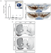
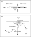
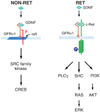
The pathways of programmed cell death (PCD) are now understood in extraordinary detail at the molecular level. Although much evidence suggests that they are likely to play a role in Parkinson's disease (PD), the precise nature of that role remains unknown. Two pathways of cell death that are especially well characterized are cyclin-dependent kinase 5-mediated phosphorylation of myocyte enhancer factor 2 and the mitogen-activated protein kinase signalling cascade. Although blockade of these pathways in animals has achieved a truly remarkable degree of neuroprotection of the neuron cell soma, it has not achieved protection of axons. Thus, there is a need to explore beyond the canonical pathways of PCD and investigate mechanisms of axon destruction. We also need to move beyond the narrow classic concept that the mechanisms of PCD are activated exclusively 'downstream', following cellular injury. Studies in the genetics of PD suggest that in some forms of the disease, activation may be an early 'upstream' event. Additionally, recent observations suggest that cell death in some contexts may not be initiated by injury, but instead by a failure of intrinsic cell survival signalling. These new points of view offer new opportunities for molecular targeting.
2010 Elsevier B.V. All rights reserved.
Figures


References
-
- Ahuja HS, Zhu Y, Zakeri Z. Association of cyclin-dependent kinase 5 and its activator p35 with apoptotic cell death. Developmental Genetics. 1997;21:258–267. - PubMed
-
- Airaksinen MS, Saarma M. The GDNF family: signalling, biological functions and therapeutic value. Nature Reviews Neuroscience. 2002;3:383–394. - PubMed
-
- Araki T, Sasaki Y, Milbrandt J. Increased nuclear NAD biosynthesis and SIRT1 activation prevent axonal degeneration. Science. 2004;305:1010–1013. - PubMed
-
- Behrens A, Sibilia M, Wagner EF. Amino-terminal phosphorylation of c-Jun regulates stress-induced apoptosis and cellular proliferation. Nature Genetics. 1999;21:326–329. - PubMed
Publication types
MeSH terms
Substances
Grants and funding
LinkOut - more resources
Full Text Sources
Medical
Miscellaneous

