. 2010:183:209-33.
doi: 10.1016/S0079-6123(10)83011-0.

Maladaptive striatal plasticity in L-DOPA-induced dyskinesia

Affiliations

Maladaptive striatal plasticity in L-DOPA-induced dyskinesia

M Angela Cenci et al. Prog Brain Res. 2010.

Abstract

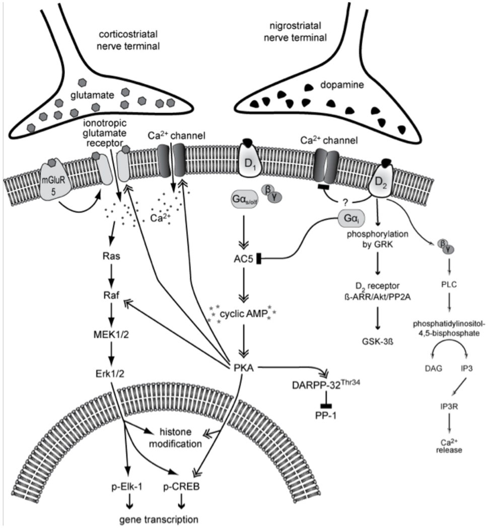
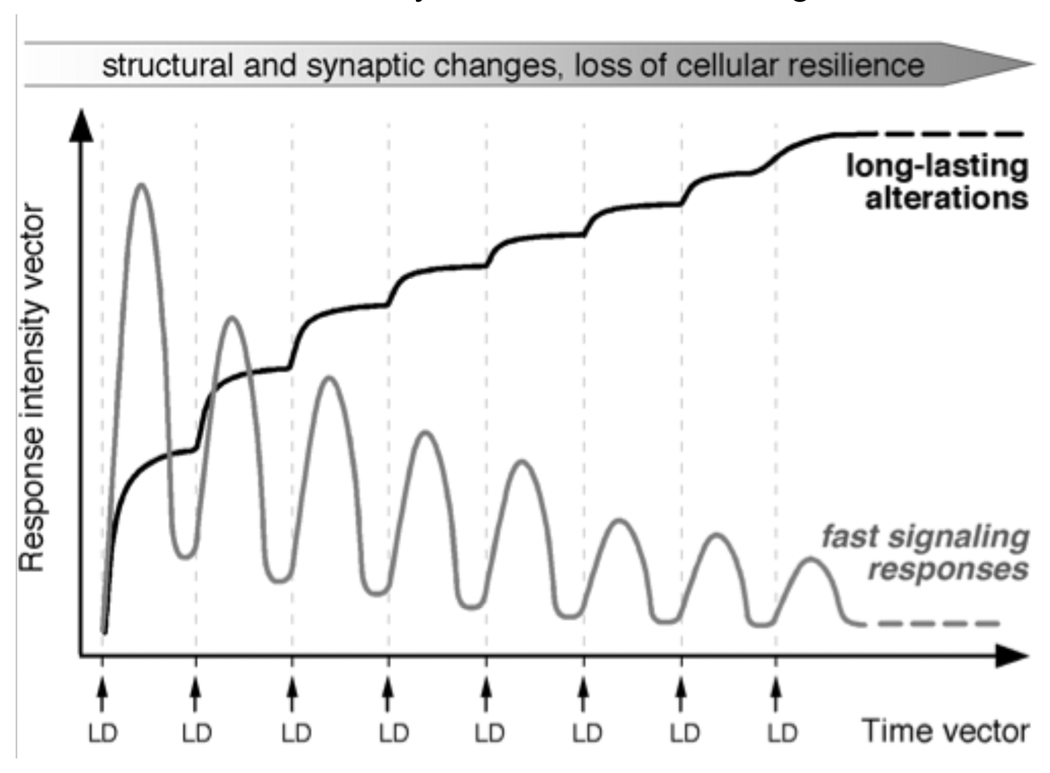
Dopamine (DA) replacement therapy with l-DOPA remains the most effective treatment for Parkinson's disease, but causes dyskinesia (abnormal involuntary movements) in the vast majority of the patients. The basic mechanisms of l-DOPA-induced dyskinesia (LID) have become the object of intense research focusing on neurochemical and molecular adaptations in the striatum. Here we review this vast literature and highlight trends that converge into a unifying pathophysiological interpretation. We propose that the core molecular alteration of striatal neurons in LID consists in an inability to turn down supersensitive signaling responses downstream of DA D1 receptors (where supersensitivity is primarily caused by DA denervation). The sustained activation of intracellular signaling pathways induced by each dose of l-DOPA leads to abnormal cellular plasticity and high bioenergetic expenditure. The over-exploitation of signaling pathways and energy reserves during treatment impairs the ability of striatal neurons to dynamically gate cortically driven motor commands. LID thus exemplifies a disorder where 'too much' molecular plasticity leads to plasticity failure in the striatum.


Figures

Figure 1
Figure 1. Canonical and non-canonical signaling cascades downstream of D1 and D2 dopamine receptors
Being coupled to stimulatory (Gs/olf) and inhibitory (Gi/o) GTP-binding proteins, D1 and D2 receptors have opposite effects on the adenylyl cyclase/cAMP/PKA/DARPP-32 cascade (‘canonical pathway’), which regulates the levels of phosphorylation of multiple cellular and nuclear targets. In particular, the cAMP/PKA/DARPP-32 pathway modulates the activity of MAPK-dependent signaling pathways downstream of glutamate receptors. It has been recently recognized that cAMP-independent pathways are also recruited following D2 receptor stimulation (non-canonical pathways; cf. Section ‘DA receptor signaling in the neurons of the intact striatum: modulation of cAMP/PKA pathways and non-canonical signaling’). Full names of the molecules shown in this drawing are given in the list of abbreviations.
Figure 2
Figure 2. Deficient densensitization of DA receptor-dependent signaling in the dyskinetic striatum
Acute treatment with l-DOPA results in a supersensitive activation of cAMP- and MAPK-dependent pathways signaling to the nucleus, here exemplified by the phosphorylation of ERK1/2 (on Thr202-Tyr204), MSK-1 (on Ser376) and by a modification of histone 3 that has been linked to chromatin remodelling during active gene transcription (phospho[Ser10]-Acetyl[Lys14]-H3). Chronic l-DOPA administration normalizes these supersensitive responses only in animals that remain free from dyskinesia during the treatment (non-dyskinetic group). Levels of signaling pathway activation remain significantly elevated above control values in dyskinetic animals. Thus, a shift towards normal molecular responses during chronic l-DOPA treatment is associated with a resistance to dyskinesia, while persistent supersensitivity is a correlate of LID. These data show both published and unpublished results obtained from 6-OHDA-lesioned rats, which received chronic (10–12 days) or acute treatment with l-DOPA (l-DOPA methyl ester, 10 mg/kg/dose combined with 15 mg/kg/dose of benserazide), or physiological saline, and were killed 30 minutes post injection. Data in (a) and (b) represent counts of immunoreactive neurons in the lateral part of the DA-denervated caudate–putamen, from animals reported in Westin et al. (2007), and five chronically l-DOPA-treated non-dyskinetic rats from the same study (not reported in the paper). Data in (c) show results from Western immunoblotting analysis. The optical density on specific immunoreactive bands was normalized to the corresponding β-actin bands, and results from each group were expressed as a percentage of saline control values. A Western blot showing bands immunoreactive for phospho[Ser10]-Acetyl[Lys14]-histone 3, total histone 3 and beta-actin is shown in (d). Full descriptions of the experimental procedures can be found in Westin et al. (2007) (for a, b) and Schroeder et al. (2008) (for c, d). Abbreviations in (d): s, saline control; a, acute l-DOPA; d, chronic l-DOPA/dyskinesia; nd, chronic l-DOPA/no dyskinesia). Values indicate group means ± SEM from 3 to 10 animals per group, which were statistically compared using one-factor analysis of variance and post-hoc Newman– Keuls test. P < 0.05 vs. *, saline-treated 6-OHDA-lesioned controls; °, acute l-DOPA; #, chronic l-DOPA with dyskinesia.
Figure 3
Figure 3. Levels of FosB/FosB-like immunoreactivity and prodynorphin mRNA are persistently upregulated in the dyskinetic striatum
(a) ΔFosB-like proteins have very slow elimination kinetics and can therefore accumulate in striatal neurons during the course of chronic l-DOPA treatment. This effect is seen only in animals that develop dyskinesia. Acute l-DOPA treatment also induces the expression of FosB/FosB-like immunoreactivity and prodynorphin mRNA, but this upregulation is transient (Cenci et al., 1999). In the experiments shown here, acutely and chronically l-DOPA-treated animals were killed at 3 hours or 2 days post-injection to better separate the effects of the chronic treatment from those of the last drug injection. (b) Prodynorphin mRNA shows the same expression pattern as FosB/ΔFosB like immunoreactivity (indeed, ΔFosB-like transcription factors mediate the upregulation of prodynorphin mRNA induced by l-DOPA in the DA-denervated striatum; see Section ‘Altered expression and regulation of transcription factors’). Values in (a) represent numbers of FosB/ΔFosB-immunoreactive cells/mm2 measured in the lateral part of the DA-denervated caudate–putamen from animals in previously published studies (Cenci et al., 1999] and Westin et al., 2007]). Data in (b) represent the hybridization signal to prodynorphin mRNA in the DA-denervated caudate–putamen (expressed as a percentage of the values on the contralateral intact side in each group) (data from Cenci et al., 1998] and Mela et al., 2007]). In each data set, values indicate group means ± SEM from 3 to 10 animals per group, P < 0.05 vs. saline-treated 6-OHDA-lesioned controls; # chronic l-DOPA with dyskinesia. Acutely and chronically l-DOPA-treated rats were compared with their own saline control group. (c–e) Cellular levels of FosB/ΔFosB immunostaining are larger in chronically l-DOPA-treated dyskinetic rats compared to acutely l-DOPA-treated animals, and a similar pattern of group differences applies to prodynorphin mRNA (f–h). Photomicrographs in (f–h) show emulsion-coated autoradiographs of striatal sections visualized under dark field optics. The sections had been hybridized with a 35S-labeled oligonucleotide probe complementary to prodynorphin mRNA. Scale bar, 50 µm. Full descriptions of all the experimental procedures can be found in our original publications.
Figure 4
Figure 4. Maladaptive plasticity of striatal neurons in LID
In animal models of LID, each dose of l-DOPA (LD) causes an abnormally large and prolonged activation of cAMP- and MAPK-dependent signaling pathways in striatal neurons (fast signaling responses). Although these responses would tend to normalize during chronic drug treatment, desensitization processes are inefficient in dyskinetic subjects. Hence, in these subjects, treatment with l-DOPA disrupts the dynamics of signaling networks that are normally under tight spatiotemporal control. Signalling dysregulation has both acute electrophysiological effects and long-term consequences on the function of striatal neurons. Long-term cellular alterations associated with LID have been documented to occur in D1/prodynorphin-positive MSN, including altered regulation of CRE/AP-1-dependent transcription, increased expression of neurotransmitter-related genes and increased synthesis and phosphorylation of cytoskeletal proteins (see Sections ‘Altered expression and regulation of transcription factors’ and ‘Potential consequences of dysregulated D1 receptor-dependent signaling’). This pattern of responses would predict the occurrence of profound structural and synaptic rearrangements in these neurons, associated with a large bioenergetic expenditure. Plastic modifications affecting D2/prepronkephalin MSN in LID are far less understood and will need to be addressed in future studies.

References

    1. Ahmed MR, Berthet A, Bychkov E, Porras G, Li Q, Bioulac BH, et al. Lentiviral overexpression of GRK6 alleviates L-dopa-induced dyskinesia in experimental Parkinson's disease. Sci Transl Med. 2010;2(28) 28ra28. - PMC - PubMed
    1. Alonso-Frech F, Zamarbide I, Alegre M, Rodríguez-Oroz MC, Guridi J, Manrique M, Valencia M, Artieda J, Obeso JA. Slow oscillatory activity and levodopa-induced dyskinesias in Parkinson's disease. Brain. 2006;129(Pt7):1748–1757. - PubMed
    1. Andersson M, Hilbertson A, Cenci MA. Striatal fosB expression is causally linked with l-DOPA-induced abnormal involuntary movements and the associated upregulation of striatal prodynorphin mRNA in a rat model of Parkinson's disease. Neurobiol Dis. 1999;6(6):461–474. - PubMed
    1. Andersson M, Konradi C, Cenci MA. cAMP response element-binding protein is required for dopamine-dependent gene expression in the intact but not the dopamine-denervated striatum. J Neurosci. 2001;21(24):9930–9943. - PMC - PubMed
    1. Andersson M, Westin JE, Cenci MA. Time course of striatal DeltaFosB-like immunoreactivity and prodynorphin mRNA levels after discontinuation of chronic dopaminomimetic treatment. Eur J Neurosci. 2003;17(3):661–666. - PubMed

Publication types

MeSH terms

LinkOut - more resources