Comparative Study
. 2010 Aug 12;67(3):373-80.
doi: 10.1016/j.neuron.2010.07.004.

The role of the TRP channel NompC in Drosophila larval and adult locomotion

Affiliations
Comparative Study

The role of the TRP channel NompC in Drosophila larval and adult locomotion

Li E Cheng et al. Neuron. .

Abstract

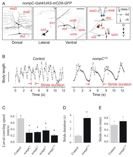
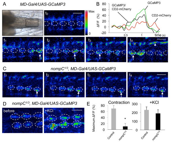
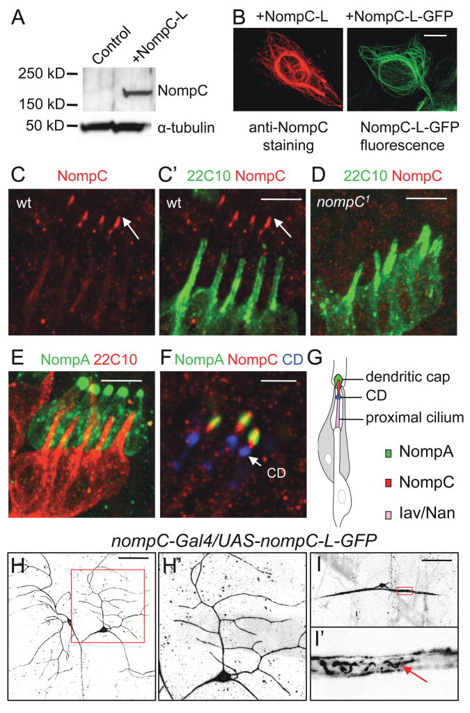
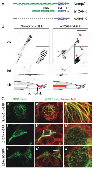
The generation of coordinated body movements relies on sensory feedback from mechanosensitive proprioceptors. We have found that the proper function of NompC, a putative mechanosensitive TRP channel, is not only required for fly locomotion, but also crucial for larval crawling. Calcium imaging revealed that NompC is required for the activation of two subtypes of sensory neurons during peristaltic muscle contractions. Having isolated a full-length nompC cDNA with a protein coding sequence larger than previously predicted, we demonstrate its function by rescuing locomotion defects in nompC mutants, and further show that antibodies against the extended C terminus recognize NompC in chordotonal ciliary tips. Moreover, we show that the ankyrin repeats in NompC are required for proper localization and function of NompC in vivo and are required for association of NompC with microtubules. Taken together, our findings suggest that NompC mediates proprioception in locomotion and support its role as a mechanosensitive channel.


Figures

Figure 1
Figure 1. NompC is expressed in larval sensory neurons and controls the pace of larval crawling
(A) NompC expression examined in nompC-Gal4/UAS-mCD8-GFP larvae. Expression was detected in class I da neurons (ddaD, ddaE, vpda), bd neurons (dbd, vbd) and chordotonal (ch) organs (lch1, lch5, vchA, vchB) in the abdominal segments. Scale bar, 50 μm. (B) Plot of body length (mm) over time (s) during peristaltic contractions for control and nompC1/3 larvae. Scale bar, 2 mm. (C–E) Quantification of crawling defects in nompC mutant larvae. Crawling speed is the length of a path (mm) divided by the time (s) it takes for the larva to complete the path. Stride duration is the average time for a larva to complete a single peristaltic contraction cycle. Stride size is the length of a path (mm) divided by the number of strides. Error bars represent SEM, n≥20. * P<0.01. See also Figure S1 and Movie S1.
Figure 2
Figure 2. NompC is required for the activation of MD neurons during peristaltic muscle contractions
(A) Ca2+ response of MD neurons to peristaltic muscle contractions in control larvae. Top left panel, a transmitted light image showing the ventral nerve cord in a larval preparation. Anterior is to the left and posterior to the right. (t0 to t4) Time-lapse images of Ca2+ signals (arrows) generated by spontaneous muscle contractions. The dorsalmedial neuropil is outlined with a dashed circle. The nerve bundles are marked with arrowheads. Color scale indicates activation level (red is the highest). Scale bar, 30 μm. (B) Representative fluorescence change (ΔF/F) of GCaMP3 and CD2-mCherry (internal reference control) in control larvae. (C) Ca2+ response to muscle contractions in nompC1/3 mutant larvae. Scale bar, 30 μm. (D) Representative images before and after adding KCl to nompC1/3 mutant larvae. Scale bar, 50 μm. (E) Quantification of GCaMP responses. Left panel, responses to muscle contractions for control (n=8) and nompC1/3 larvae (n=10). Right panel, responses to KCl for control (n=3) and nompC1/3 larvae (n=3). The GCaMP signal is normalized to CD2-mCherry reference signal. * p<0.01. See also Movie S2.
Figure 3
Figure 3. A full-length nompC cDNA rescues the locomotion defects in nompC1/3 mutant larvae and adult flies
(A) The exon-intron structures of nompC-L and nompC-A aligned with the genomic sequence. Exons are indicated by shaded boxes/triangles and introns by dotted lines. (B, C) Quantification of larval crawling speed and stride duration with the expression of NompC-L or NompC-L-GFP by different Gal4 drivers. SN, sensory neurons; MD, multi-dendritic neurons; bd/I, bd and class I da neurons. (D) Quantification of adult fly walking speed with NompC-L expression. (E) Quantification of stride duration with expression of the truncated NompC, Δ12ANK-GFP and Δ29ANK-GFP. Error bars represent SEM, n≥20. * P<0.01. See also Figure S2, Movie S3 and Table S1.
Figure 4
Figure 4. NompC distribution in embryonic and larval sensory neurons
(A) Western blots with the NompC antibody in NompC-L-transfected HEK 293 cells. (B) Immunostaining with the NompC antibody in NompC-L-transfected cells and GFP fluorescence in NompC-L-GFP-transfected cells. Scale bar, 10 μm. (C, D) Co-staining of chordotonal organs (lch5) with the NompC antibody (red) and neural-specific cytoskeletal maker mAb 22C10 (green) in wild-type and nompC1 mutant embryos. Scale bar, 5 μm. (E) In embryos expressing GFP-NompA, co-staining of GFP (green) and mAb 22C10 (red) marks the NompA-enriched dendritic caps. Scale bar, 5 μm. (F) Co-staining of embryos expressing GFP-NompA (green) with mAb 21A6/anti-EYS (blue), a marker for the ciliary dilation (CD), and the NompC antibody (red). Scale bar, 3 μm. (G) A schematic of a single chordotonal organ showing the localization of NompA in dendritic caps, NompC in distal ciliary tips and Iav/Nan in the proximal cilium. (H, I) Localization of NompC-L-GFP in larval class I da neurons (H) and bd neurons (I). The arrow indicates NompC-L-GFP fluorescence in filamentous structures within the proximal dendrites. The boxed area in H and I is enlarged and shown in H′ and I′, respectively. Scale bar, 50 μm. See also Figure S3.
Figure 5
Figure 5. Ankyrin repeats of NompC are required for ciliary localization and microtubule association
(A) A schematic of full-length and truncated NompC-L protein structures. ANK, ankyrin domain; TM, transmembrane domain; TRP, TRP box domain. (B) Localization of NompC-L-GFP and Δ12ANK-GFP in chordotonal organs (upper panels) and bd neurons (middle panels). The lch5 sensory neurons are indicated by *, the base of the cilium by an arrowhead and the ciliary tip by an arrow. The bd axon is indicated by an arrow. A schematic interpretation of the localization of NompC-L-GFP and Δ12ANK-GFP in chordotonal organs are shown in the bottom panels. pc, proximal cilium; cd, ciliary dilation; dc, distal ciliary tip. Scale bars, 20 μm. (C) Localization of NompC-L-GFP, Δ12ANK-GFP and Δ29ANK-GFP in HEK 293 cells. Full-length and truncated NompC-L protein was visualized by the GFP fluorescence (green). The cells were co-stained with anti-α-tubulin (red). Boxed area in a′, b′, c′ is enlarged and shown in a″, b″, c″ respectively. Scale bar, 10 μm. See also Figure S4.

Comment in

References

    1. Ainsley JA, Pettus JM, Bosenko D, Gerstein CE, Zinkevich N, Anderson MG, Adams CM, Welsh MJ, Johnson WA. Enhanced locomotion caused by loss of the Drosophila DEG/ENaC protein Pickpocket1. Curr Biol. 2003;13:1557–1563. - PubMed
    1. Albert JT, Nadrowski B, Gopfert MC. Mechanical signatures of transducer gating in the Drosophila ear. Curr Biol. 2007;17:1000–1006. - PubMed
    1. Bodmer R, Jan YL. Morphological differentiation of the embryonic peripheral neurons in Drosophila. Roux’s Archives of Developmental Biology. 1987;196:69–77. - PubMed
    1. Chung YD, Zhu J, Han Y, Kernan MJ. nompA encodes a PNS-specific, ZP domain protein required to connect mechanosensory dendrites to sensory structures. Neuron. 2001;29:415–428. - PubMed
    1. Ernstrom GG, Chalfie M. Genetics of sensory mechanotransduction. Annu Rev Genet. 2002;36:411–453. - PubMed

Publication types

Associated data