Comparative Study
. 2010 Aug 12;67(3):381-91.
doi: 10.1016/j.neuron.2010.06.032.

C. elegans TRP family protein TRP-4 is a pore-forming subunit of a native mechanotransduction channel

Affiliations
Comparative Study

C. elegans TRP family protein TRP-4 is a pore-forming subunit of a native mechanotransduction channel

Lijun Kang et al. Neuron. .

Abstract

Mechanotransduction channels mediate several common sensory modalities such as hearing, touch, and proprioception; however, very little is known about the molecular identities of these channels. Many TRP family channels have been implicated in mechanosensation, but none have been demonstrated to form a mechanotransduction channel, raising the question of whether TRP proteins simply play indirect roles in mechanosensation. Using Caenorhabditis elegans as a model, here we have recorded a mechanosensitive conductance in a ciliated mechanosensory neuron in vivo. This conductance develops very rapidly upon mechanical stimulation with its latency and activation time constant reaching the range of microseconds, consistent with mechanical gating of the conductance. TRP-4, a TRPN (NOMPC) subfamily channel, is required for this conductance. Importantly, point mutations in the predicted pore region of TRP-4 alter the ion selectivity of the conductance. These results indicate that TRP-4 functions as an essential pore-forming subunit of a native mechanotransduction channel.


Figures

Figure 1
Figure 1. In vivo patch-clamp recording of mechanoreceptor currents (MRCs) in the dopamine neuron CEP
(A) A schematic illustrating the morphology of CEP (Perkins et al., 1986). A glass probe (2 μm in diameter) driven by a piezo actuator was used to deliver mechanical stimuli towards the CEP cilium. (B) MRCs in CEP evoked by a 4 μm stimulus. CEP cell body was voltage-clamped at −75 mV. The dotted line in this trace and all other traces in this paper denotes the baseline (zero current). Most, if not all, worm neurons particularly head sensory neurons are known to be nearly isopotential (voltage is quite uniform throughout the neuron), and thus voltage attenuation (space clamp) is minimal (Goodman et al., 1998). (C) I-V relations of MRCs in CEP. Peak current values were used here and throughout the paper. n=6.
Figure 2
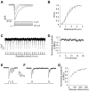
Figure 2. The amplitude of MRCs is stimulus-strength dependent and they do not desensitize to repetitive stimuli with long intervals
(A-B) The amplitude of MRCs is stimulus-strength dependent. Mechanical stimuli of varying displacement were used to stimulate CEP. Shown in (A) are sample traces recorded from a CEP neuron in response to 0.5, 1, 2, 3, and 4 μm of displacement. Each trace was averaged from 4 sweeps. I/Imax values were plotted against displacement in (B), and the data were fit with a Boltzmann function: I/Imax = 1/(1 + exp(−(XX1/2)/Xslope)), where I denotes current amplitude and X represents displacement. n=6. (C-D) MRCs in CEP do not desensitize to repetitive stimuli with long intervals. Repetitive stimuli (4 μm) were applied to CEP neurons for 50 times with an interval of 2 sec between each stimulus. Shown in (C) is a sample trace of the first 20 responses, and the data are summarized in (D). n=9. (E-G) MRCs in CEP show desensitization to repetitive stimuli with short intervals. (E) Two consecutive stimuli (4 μm) with a short interval (30 ms) were applied to CEP. The amplitude of MRCs evoked by the second stimulus was greatly reduced compared to that triggered by the first stimulus, indicating desensitization. (F) However, no such desensitization was observed in CEP when we applied repetitive stimuli with a long interval (300 ms). Traces shown in (E) and (F) were from the same neuron. (G) Data summary. n=4. Error bars represent SEM.
Figure 3
Figure 3. MRCs in CEP show rapid activation kinetics and short latency
(A) The activation kinetics of MRCs becomes faster with increasing stimulus strength, reaching the micro-seconds range. The activation time constants of MRCs were plotted as a function of displacement, and the solid line represents a single exponential fit to the data. n=6. (B-C) The latency of MRCs reduces with increasing stimulus strength, reaching the micro-seconds range. Shown in (B) are sample traces, and the arrow indicates the onset of the stimulus. Latency values were plotted against displacement in (C), and the solid line represents a single exponential fit to the data. n=6.
Figure 4
Figure 4. Non-stationary noise analysis
(A-B) Non-stationary noise analysis of MRCs. Shown in (A) are the ensemble average MRC trace (top) and the variance trace (bottom) from a CEP neuron in response to 50 repetitive stimuli (1 μm) with an interval of 2 sec between each stimulus, a condition under which desensitization to repetitive stimuli did not occur (Figure 2C-D). The variance values were plotted against mean current, and the data were fit with the equation: δ2(I)= iI - I2/N, where δ2 is the variance, i represents the single channel current, and N indicates the number of channels available for activation (see experimental procedures for details). For the cell shown in (B), i = 1.2 pA and N = 23.
Figure 5
Figure 5. Adaptation extends the dynamic range of CEP mechanosensitivity
(A-B) Adaptation extends the dynamic range of response in CEP. Shown in (A) is a sample trace of a CEP neuron in response to a series of testing and adapting stimuli (1μm). The I/Imax values under different adapting stimulus strength were plotted against displacement, and the data were fit with a Boltzmann function: I/Imax = 1/(1 + exp(−((X − X0) − X1/2)/Xslope)), where I denotes current amplitude, X represents displacement, and X0 indicates adapting displacement. The actual shift of the stimulus-response curve estimated based on ΔX1/2 is: 1.0 μm and 2.4 μm under a 1.0 μm and 2.5 μm of adapting displacement, respectively. Each curve represents data points from at least 3 cells. (C) Time course of the adaptive shift of the stimulus-response curve. Unlike in (B) where testing pulses were applied at a fixed time point (200 ms) after the onset of adapting pulses of varying strength, here testing pulses were delivered at varying time points (0 to 400 ms) after the onset of a adapting pulse of fixed strength (1 μm). X1/2 value under each condition was calculated as described in (B). By doing so, the actual shift of the stimulus-response curve (i.e. ΔX1/2 values) was obtained for each condition (time point), and this value was plotted against time points as shown in (C). n=7.
Figure 6
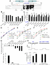
Figure 6. TRP-4 is required for MRCs in CEP
(A-B) TRP-4 is required for MRCs in CEP. (A) wild-type. (B) No MRC was detected in CEP of trp-4(sy695) mutant worms in response to a saturating stimulus (4 μm). sy695 is a null allele of trp-4 (Li et al., 2006). (C) Transgenic expression of wild-type trp-4 cDNA in CEP under the dopamine neuron-specific promoter dat-1 (Lints and Emmons, 1999) rescues MRCs in trp-4(sy695) mutant worms. (D) Bar graph summarizing the data in (A-C). ocr-2 cDNA under the dat-1 promoter failed to rescue MRCs in CEP. n≥6. (E) trp-4 mutant worms are defective in the basal slowing behavioral response, and this phenotype can be rescued by transgenic expression of wild-type trp-4 cDNA, but not ocr-2 cDNA. ocr-2 cDNA can rescue the osmotic and octanol avoidance defects of ocr-2 mutants (see Figure S2D-E). n≥10. *p<0.05 (t test). (F) I-V relations of CEP MRCs recorded from rescued worms. n=7. (G) I-V relations of CEP MRCs recorded from rescued worms and wild-type worms are nearly identical.
Figure 7
Figure 7. TRP-4 is an essential pore-forming subunit of the mechanotransduction channel in CEP
(A) Sequence alignment of the putative pore region of TRP-4 and its homologues. The bracket denotes the putative pore helix that is predicted to form an alpha helix structure by the program PSIPRED. Other programs (e.g. Prof and SSpro) also yield a similar prediction. The double-headed arrow indicates the putative selectivity filter. The sequences outside the pore region but within the S5-S6 linker area are not shown for other organisms as they do not show much homology. Asterisks in red mark the negatively charged residues D and E that were mutated in various TRP-4 mutant forms. TRP-4 homologues are found in Xenopus, zebrafish and Drosophila but not higher vertebrates. (B) MRC amplitude in CEP recorded from transgenic worms expressing various mutant forms of TRP-4. Wild-type TRP-4 rescue data from 6D is also included. All transgenes were expressed in the trp-4(sy695) mutant background. It should be noted that the expression level of different transgenes may vary and such variation may contribute to the difference in the amplitude of MRCs carried by different forms of TRP-4. Displacement: 4 μm. n≥5. (C) Rescue of the basal slowing behavioral response by transgenes encoding various mutant forms of TRP-4. All transgenes were expressed in the trp-4(sy695) mutant background. n≥10. *p<0.05 (t test). (D-F) I-V relations. Wild-type TRP-4 was included in each panel for caparison. (G) Summary of reversal potential data. Wild type TRP-4 and various mutant forms of TRP-4 were all expressed in the trp-4(sy695) mutant background. Data from wild type worms was also shown for comparison. n≥5. (H-I) The “pore-dead” mutant EPD1739-41KPK blocks endogenous MRCs in CEP of wild-type worms. The tiny remaining current may result from those very few homomeric wild-type TRP-4 channels that did not heteromerize with the mutant, assuming that TRP-4 functions as a tetramer. Wild-type data from 6D is also included. Displacement: 4 μm. n≥6. (J) Wild-type worms expressing the “pore-dead” mutant form EPD1739-41KPK lack the basal slowing behavioral response. n≥12. *p<0.05 (t test).

Comment in

References

    1. Bautista DM, Jordt SE, Nikai T, Tsuruda PR, Read AJ, Poblete J, Yamoah EN, Basbaum AI, Julius D. TRPA1 mediates the inflammatory actions of environmental irritants and proalgesic agents. Cell. 2006;124:1269–1282. - PubMed
    1. Bounoutas A, Chalfie M. Touch sensitivity in Caenorhabditis elegans. Pflugers Arch. 2007;454:691–702. - PubMed
    1. Brockie PJ, Mellem JE, Hills T, Madsen DM, Maricq AV. The C. elegans glutamate receptor subunit NMR-1 is required for slow NMDA-activated currents that regulate reversal frequency during locomotion. Neuron. 2001;31:617–630. - PubMed
    1. Christensen AP, Corey DP. TRP channels in mechanosensation: direct or indirect activation? Nature reviews. 2007;8:510–521. - PubMed
    1. Colbert HA, Smith TL, Bargmann CI. OSM-9, a novel protein with structural similarity to channels, is required for olfaction, mechanosensation, and olfactory adaptation in Caenorhabditis elegans. J Neurosci. 1997;17:8259–8269. - PMC - PubMed

Publication types

MeSH terms

Substances