. 2010 Aug 11;18(8):955-65.
doi: 10.1016/j.str.2010.04.017.

Molecular basis for the association of human E4B U box ubiquitin ligase with E2-conjugating enzymes UbcH5c and Ubc4

Affiliations

Molecular basis for the association of human E4B U box ubiquitin ligase with E2-conjugating enzymes UbcH5c and Ubc4

Robert C Benirschke et al. Structure. .

Abstract

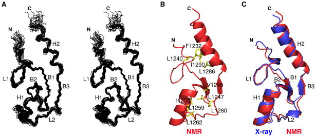
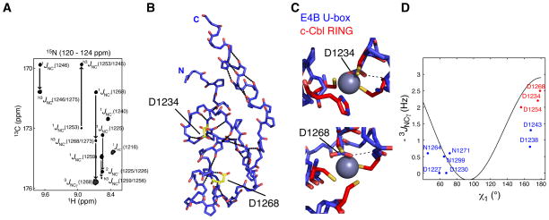
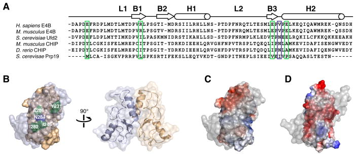
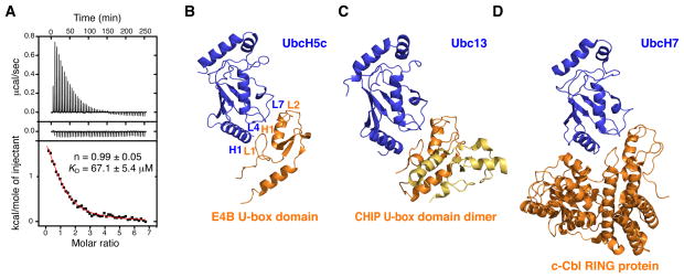
Human E4B, also called UFD2a, is a U box-containing protein that functions as an E3 ubiquitin ligase and an E4 polyubiquitin chain elongation factor. E4B is thought to participate in the proteasomal degradation of misfolded or damaged proteins through association with chaperones. The U box domain is an anchor site for E2 ubiquitin-conjugating enzymes, but little is known of the binding mechanism. Using X-ray crystallography and NMR spectroscopy, we determined the structures of E4B U box free and bound to UbcH5c and Ubc4 E2s. Whereas previously characterized U box domains are homodimeric, we show that E4B U box is a monomer stabilized by a network of hydrogen bonds identified from scalar coupling measurements. These structural studies, complemented by calorimetry- and NMR-based binding assays, suggest an allosteric regulation of UbcH5c and Ubc4 by E4B U box and provide a molecular basis to understand how the ubiquitylation machinery involving E4B assembles.


Figures

Figure 1
Figure 1. Solution and Crystal Structures of E4B U-box
(A) Superposition of the 20 lowest energy NMR structures of E4B U-box in stereoview showing only the N, Cα , and C’ trace for aa 1228 to 1300. The α-helices (H), β-strands (B) and loops (L) are indicated. (B) Ribbon representation of the NMR structure of E4B U-box showing only residues 1226 to 1302 and amino acid side chains (yellow) forming two hydrophobic cores. (C) Overlay of the crystal (aa 1226 to 1300) and NMR (aa 1226 to 1300) structures of E4B U-box where α-helices (H), β-strands (B) and loops (L) are indicated. See also Figure S1. Structure representations were prepared using MOLMOL (Koradi et al., 1996) or PyMOL (http://pymol.sourceforge.net/).
Figure 2
Figure 2. Identification of Hydrogen Bonds in E4B U-box by NMR Spectroscopy
(A) A region from the constant time 3D J-HNCO NMR spectrum of E4B U-box showing peaks correlated through 1JNC, 2JNC, 3JNCγ and h3JNC’ scalar couplings. (B) The nineteen H-bonds in E4B U-box that are listed in Table S3 were identified through h3JNC’ scalar coupling measurements. The dotted lines illustrate these H-bonds. Residues Asp1232 and Asp1268 for which side chain h3JNC’ scalar couplings were measured are shown. Asp1232 and Asp1268 correspond to the zinc binding regions of RING domains. (C) Asp1232 and Asp1268 of E4B U-box approximately occupy the zinc centers (purple spheres) in c-Cbl RING domain (PDB entry 1FBV) when the U-box and RING domains are superimposed. H-bonds involving the two aspartate side chains were determined from h3JNC’ scalar coupling measurements and are shown by the dashed lines. (D) Calibration curve for the dihedral angle (χ1) dependence of 3JNCγ scalar couplings in aspartate and asparagine residues. Blue and red points correlate experimental 3JNCγ and corresponding χ1 angle values from the crystal structure of E4B U-box. The red points correspond to residues for which a side chain carbonyl participates in a H-bond as determined from h3JNC scalar couplings. See also Table S1 for the values of measured coupling constants.
Figure 3
Figure 3. Electrostatic Surface Potentials of E4B and CHIP U-box Domains Affect Oligomerization States
(A) Sequence alignment of U-box domains from different E3 ubiquitin ligases indicating locations of secondary structure elements. CHIP and Prp19 are dimers while Ufd2 and E4B are monomers. Key CHIP and Prp19 residues mediating homodimerization are highlighted. Hydrophobic residues in CHIP and Prp19 (green box) correspond to charged residues in E4B while an asparagine residue (Asn284) in mouse CHIP (blue box) is replaced by a valine residue (Val1283) in human E4B. (B) Molecular representation of mouse CHIP homodimer showing the important dimerization interface residues described in A. (C) Electrostatic surface potential of mouse CHIP U-box homodimer (PDB entry 2C2V) showing predominantly non-polar character at the dimerization interface. Foremost protomer is rendered transparent for clarity. (D) Electrostatic surface potential of a hypothetical homodimer of E4B U-box showing a negatively charged dimer interface.
Figure 4
Figure 4. Interaction of E4B U-box with UbcH5c and Ubc4 E2 Conjugating Enzymes
(A) Isothermal titration calorimetry of UbcH5c with E4B U-box. Shown are the integrated heat measurements from injecting 3 mM E4B U-box into the calorimeter cell containing UbcH5c at an initial concentration of 100 μM (top panel) or buffer solution (middle panel). A standard one-site model was used for curve fitting (bottom panel) in the determination of KD and stoichiometry (n), the values of which are shown. (B) Crystal structure of human E4B U-box in complex with UbcH5c. (C) Crystal structure of mouse CHIP U-box in complex with Ubc13 (PDB entry 2C2V). (D) Crystal structure of the human c-Cbl RING E3 ligase in complex with UbcH7 (PDB entry 1FBV). See also Figure S2.
Figure 5
Figure 5. Interaction Interface Between E4B U-box and UbcH5c
(A) E4B U-box and UbcH5c interface showing possible salt bridges and hydrogen bonds. (B) E4B U-box and UbcH5c interface showing how F62 and S94-P95-A96 (S-P-A motif) of UbcH5c interact with E4B U-box. (C) E4B U-box and UbcH5c interface illustrating how L1236 and M1237 of E4B U-box interact with UbcH5c. See also Figure S3 for the electron density map of the E4B U-box–UbcH5c complex.
Figure 6
Figure 6. NMR Titrations and Mapping of Interaction Interfaces in E4B U-box, UbcH5c and Ubc4
(A, B) Superposition of the 1H-15N HSQC titration spectra of 15N-labeled E4B U-box from free (black) to the bound (red) states with nonlabeled Ubc4 (A) and UbcH5c (B). The arrows indicate the directions of the titrations. (C) In the crystal structure of E4B U-box and UbcH5c complex, E4B residues affected by UbcH5c titration are colored red (signals with chemical shift changes ≥ 0.02 ppm) or blue (weakened or disappeared signals, namely those of Asp1234, Leu1236, Asp1238, Leu1247, Ile1258, Asn1271 and Arg1272). The active site Cys85 is shown in yellow. (D, E) Superposition of the 1H-15N HSQC titration spectra of 15N-labeled Ubc4 (D) or UbcH5c (E) from the free (black) to the bound (red) states with nonlabeled E4B U-box. The arrows indicate the directions of the titrations. (F) In the crystal structure of E4B U-box and UbcH5c complex, UbcH5c residues affected by E4B U-box titration are colored red (signals with chemical shift changes from 0.03 to 0.06 ppm) or blue (chemical shift changes from 0.07 to 0.16 ppm). The active site Cys85 is shown in yellow. (G) UbcH5c residues distant from the binding interface with E4B U-box and for which changes in chemical shifts were detected are shown in gray. The active site Cys85 is shown in yellow.
Figure 7
Figure 7. Model Structure of E4B, UbcH5c and Ubiquitin Ternary Complex
The structures of budding yeast Ufd2 (aa 188-961 from PDB entry 2QIZ), human UbcH5c–ubiquitin (PDB entry 2FUH) and human E4B U-box–UbcH5c (this study) were superimposed to model the complex. Ubiquitin (yellow), UbcH5c (gray) and Ufd2 (body in green and U-box in blue) fit together in the model with very few steric clashes and leave the active site cysteine of UbcH5c (red) accessible to react with a ubiquitin molecule.

Comment in

References

    1. Adams PD, Grosse-Kunstleve RW, Hung LW, Ioerger TR, McCoy AJ, Moriarty NW, Read RJ, Sacchettini JC, Sauter NK, Terwilliger TC. PHENIX: building new software for automated crystallographic structure determination. Acta Crystallogr Sect D. 2002;58:1948–1954. - PubMed
    1. Andersen P, Kragelund BB, Olsen AN, Larsen FH, Chua NH, Poulsen FM, Skriver K. Structure and biochemical function of a prototypical Arabidopsis U-box domain. J Biol Chem. 2004;279:40053–40061. - PubMed
    1. Aravind L, Koonin EV. The U box is a modified RING finger - a common domain in ubiquitination. Curr Biol. 2000;10:132–134. - PubMed
    1. Botuyan MV, Nominé Y, Yu X, Juranić N, Macura S, Chen J, Mer G. Structural basis of BACH1 phosphopeptide recognition by BRCA1 tandem BRCT domains. Structure. 2004;12:1137–1146. - PMC - PubMed
    1. Brzovic PS, Lissounov A, Christensen DE, Hoyt DW, Klevit RE. A UbcH5/ubiquitin noncovalent complex is required for processive BRCA1-directed ubiquitination. Mol Cell. 2006;21:873–880. - PubMed

Publication types

MeSH terms

Substances

LinkOut - more resources