. 2010 Aug 9;190(3):443-60.
doi: 10.1083/jcb.201006044.

KIF17 stabilizes microtubules and contributes to epithelial morphogenesis by acting at MT plus ends with EB1 and APC

Affiliations

KIF17 stabilizes microtubules and contributes to epithelial morphogenesis by acting at MT plus ends with EB1 and APC

Fanny Jaulin et al. J Cell Biol. .

Abstract

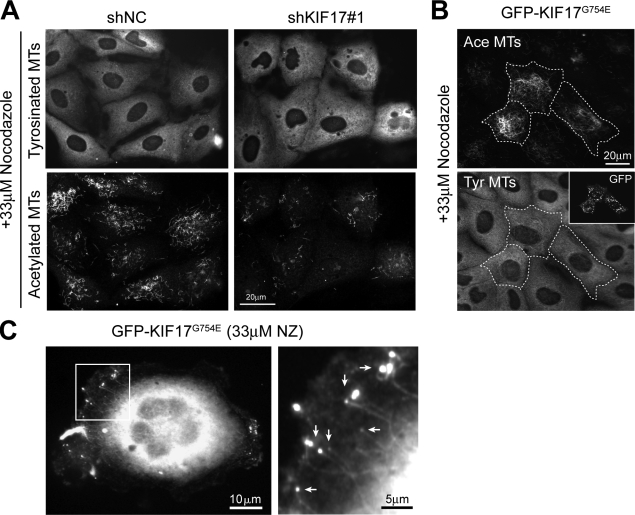
Epithelial polarization is associated with selective stabilization and reorganization of microtubule (MT) arrays. However, upstream events and downstream consequences of MT stabilization during epithelial morphogenesis are still unclear. We show that the anterograde kinesin KIF17 localizes to MT plus ends, stabilizes MTs, and affects epithelial architecture. Targeting of KIF17 to plus ends of growing MTs requires kinesin motor activity and interaction with EB1. In turn, KIF17 participates in localizing adenomatous polyposis coli (APC) to the plus ends of a subset of MTs. We found that KIF17 affects MT dynamics, polymerization rates, and MT plus end stabilization to generate posttranslationally acetylated MTs. Depletion of KIF17 from cells growing in three-dimensional matrices results in aberrant epithelial cysts that fail to generate a single central lumen and to polarize apical markers. These findings implicate KIF17 in MT stabilization events that contribute to epithelial polarization and morphogenesis.


Figures

Figure 1.
Figure 1.
KIF17 colocalizes with EB1 at MT plus ends. (A) Immunostaining of tyrosinated α-tubulin and KIF17 in Caco2 cells. Color overlay: enlargement of boxed regions showing KIF17 (green) at the plus ends of MTs (red, see arrows). (B) Immunostaining of KIF17 and EB1 in Caco2 and MDCK cells. Overlay panels show magnifications of EB1 (red) and KIF17 (green) from the boxed regions. In the bottom overlay panels, the KIF17 image was shifted 3 pixels to highlight the coincident staining pattern. (C) High-magnification images of EB1 and KIF17 in Caco2 cells. Arrows, KIF17 puncta at the head of EB1 comets; arrowheads, KIF17 puncta along the EB1 tail. (D) Quantification of KIF17 colocalization with EB1-labeled MTs in Caco2 (n = 4,575) and MDCK (n = 2,622) cells (n = the number of EB1-labeled MT ends analyzed in at least three experiments). Random colocalization was determined and subtracted from each dataset as described in Material and methods. +BP, analysis after preincubation of KIF17 IgG with immunogenic peptide (n = 1,203); Tot., total EB1-labeled MTs throughout entire cell (n = 2,622); Pro., EB1-labeled MTs in cell protrusions (n = 231). Error bars indicate SEM.
Figure 2.
Figure 2.
KIF17 motor domain interacts with EB1. (A) Immunoprecipitation of endogenous KIF17 or KIF3 from HEK-293 cells (AP, affinity purified IgG). Western blots probed for KIF17, KIF3A, or EB1. 1/20 volume used for immunoprecipitation was loaded for total lysates. At this concentration, KIF17 is not detected. (B) Schematic diagram of EB1 and KIF17 constructs. Numbers indicate amino acid position. (C) Immunoprecipitation of myc-tagged KIF17-FL, KIF17-M, or KIF17-T from MDCK cells cotransfected with GFP-tagged EB1-FL, EB1-N, or EB1-C. Immunoprecipitates and total lysates were blotted with anti-myc and anti-GFP antibodies. (D) Binding of in vitro translated HA-KIF17-FL to purified GST-tagged EB1-FL (FL), EB1-C (C), or EB1-N (N). TL, total lysate. (E) Pull-down of GFP-tagged EB1 constructs expressed in MDCK cells by purified GST-KIF17-M. (F) Coimmunoprecipitation of myc-EB1-FL and GFP-KIF17-M, GFP-KIF17-MIP, or GFP-KIF17-MΔ5 from MDCK lysates.
Figure 3.
Figure 3.
EB1 targets KIF17 to MTs. (A) Immunostaining of endogenous KIF17 and transfected myc-EB1-FL or myc-EB1-FLEE in Caco2 cells. Color overlays show boxed regions at higher magnification. (B) Line-scan analysis of the number and fluorescence intensity of KIF17 puncta along the distal 10 µM of individual MTs in cells expressing myc-EB1-FL or myc-EB1-FLEE. Data derived from three experiments in which 487 and 339 puncta along 42 MTs were analyzed in cells expressing myc-EB1 wild-type (WT) or mutant (EE) proteins. (C) Immunostaining of KIF17 and tyrosinated MTs in EB1-depleted cells. Color overlay shows an enlargement of the boxed regions. Arrows, MT ends decorated by KIF17; arrowheads, MT ends not decorated by KIF17. Western blot shows EB1 and tubulin levels in siNC or siEB1 transfected cells. Graph shows quantification of MT ends decorated by KIF17 in siNC or siEB1 cells (n = 3,058 and n = 3,882, respectively, compiled from three experiments). Error bars indicate SEM. (D) Immunostaining of tyrosinated MTs and EB1 in control and KIF17-depleted cells. Western blot shows KIF17 and glyceraldehyde 3-phosphate dehydrogenase (GAPDH) levels in Caco2 cells infected with lentiviral control (shNC) or KIF17 shRNAs (shKIF17#1 and shKIF17#2 target two unique sequences).
Figure 4.
Figure 4.
Association of GFP-KIF17G754E with MT plus ends requires kinesin ATPase and MT-binding activities. (A) Single frame from a time-lapse recording of MDCK cells expressing GFP-KIF17-FL. Inset shows a kymograph of a moving GFP-KIF17-FL puncta during 20 s of the time-lapse recording (Video 1). The inset shows an enlargement of the boxed region. (B) Alignment of the hinge regions of C. elegans OSM-3 and human KIF17. The gray box shows the position of the G-to-E mutations in KIF17 and OSM-3. Numbers at left and right indicate amino acid position. Asterisks indicate sequence identities between KIF17 and OSM-3. (C) Single frame from a time-lapse recording of Caco2 cells injected with GFP-KIF17G754E cDNA (Video 2). Boxed regions (a and b) are enlarged in the bottom panels. (B, right) Selected frames from the time-lapse recording. Asterisks indicate a KIF17 puncta moving on MTs toward the plus end. (D) Images of GFP-KIF17G754E, GFP-KIF17G754E-G243A, and GFP-KIF17G754E-R288/294A expressed by cDNA injection in live or prepermeabilized and fixed MDCK cells. GFP-KIF17G754E localizes in cell extensions at MT ends (arrows), whereas GFP-KIF17G754E-G243A and GFP-KIF17G754E-R288/294A do not (arrowheads).
Figure 5.
Figure 5.
KIF17 coiled-coil region binds APC independently of EB1. (A) Colocalization of GFP-fAPC and mRFP-KIF17G754E at plus ends of a subset of MTs in Caco2 cells 3 h after cDNA injection. The boxed region is magnified on the right. (B) Immunostaining of APC and tyrosinated tubulin in MDCK cells expressing GFP-KIF17G754E. Color overlays (a and b) show magnifications of the boxed regions. (C) Schematic of wild-type (WT) and C-terminally truncated APC present in Caco2 cells. The red arrowhead shows the EB1 binding site, which is missing in Caco2 APC. Immunoblots show endogenous APC, EB1, KIF17, and KIF3 in total lysates (TL) and immunoprecipitates from Caco2 cells incubated with rabbit IgG or anti-APC IgG. Numbers on top indicate amino acid position. (D) Immunoblots showing total lysates and immunoprecipitated APC (endogenous) and GFP-KIF17 constructs expressed in Caco2 cells.
Figure 6.
Figure 6.
KIF17 is necessary for APC localization at MT plus ends. (A) Immunostaining of endogenous APC (red) in MDCK cells fixed 1 d after transfection with GFP-KIF17-S (green). Arrows, APC clusters in cell protrusions in nonexpressing cells; arrowheads, protrusions lacking APC in a cell expressing GFP-KIF17-S. (B) Percentage of GFP-KIF17-S–expressing cells (n = 183) or surrounding controls (n = 189) with APC clusters in protrusions. Data were compiled from three experiments. Error bars indicate SEM. (C) Immunostaining of APC and MTs in MDCK cells expressing control (shNC) or KIF17 (shKIF17#1) shRNAs. Arrows, APC clusters localizing at MT ends in cell protrusions; arrowheads, cell protrusions lacking APC clusters at MT ends. Insets show enlarged views of the boxed regions. (D) Rescue of APC localization in cell protrusions by expression of GFP-KIF17G754E in KIF17-depleted MDCK cells. Boxed regions are shown at high magnification on the right.
Figure 7.
Figure 7.
KIF17 affects MT acetylation in epithelial cells. (A and B) Immunostaining of tyrosinated and acetylated tubulin in MDCK cells infected with control (shNC) or KIF17 (shKIF17#1) shRNA (A), or fixed 5 h after microinjection of GFP-KIF17G754E cDNA (B). (C) Quantitative immunoblot analysis of acetylated tubulin in MDCK cells treated with control or KIF17 shRNA, or expressing GFP or GFP-KIF17G754E. The ratio of acetylated tubulin/GAPDH was normalized to 1 in controls. Data were derived from 3–5 experiments. Error bars indicate SEM. (D) KIF17-depleted MDCK cells fixed 3 h after injection of GFP-KIF17G754E cDNA and stained for tyrosinated and acetylated MTs. Outlined regions indicate injected cells. Insets show the injected GFP-KIF17G754E.
Figure 8.
Figure 8.
KIF17 attenuates MT polymerization. (A) Maximum projection of EB1-FL-GFP in 10 sequential frames (2-s intervals) of a time-lapse recording of Caco2 cells. EB1-GFP was expressed by cDNA microinjection. The first frame is shown in blue, the last frame in red, and intermediary frames in green. (B) Analysis of EB1-GFP–labeled MT dynamics (persistent growth) and mean polymerization rates (net displacement/time) in Caco2 cells expressing control (shNC) or KIF17 (shKIF17#1) shRNAs and in cells expressing RFP or RFP-KIF17G754E. Data compiled from at least five cells/experiment in three experiments. n = the number of EB1-labeled MTs analyzed. (C–E) Histograms show polymerization rates of EB1-GFP–labeled MTs in Caco2 cells infected with shNC or shKIF17#1 (C), coexpressing RFP or RFP-KIF17G754E (D), or expressing EB1-FL-GFP or EB1-FLEE-GFP (E). Data were compiled from at least six cells per experiment in three experiments. n = the number of EB1-labeled MT ends analyzed.
Figure 9.
Figure 9.
KIF17 affects MT stabilization in epithelial cells. (A) Immunostaining of tyrosinated and acetylated tubulin in MDCK cells infected with shNC or shKIF17#1 and treated for 45 min with 33 µM NZ. (B) Acetylated and tyrosinated tubulin in MDCK cells (outlined cells and inset), treated for 1 h with 33 µM NZ. (C) Caco2 cells injected with GFP-KIF17G754E cDNA and treated for 15 min with 33 µM NZ. An enlargement of the boxed region is shown on the right. Arrows point to GFP-KIF17G754E at the ends of NZ-resistant MTs.
Figure 10.
Figure 10.
KIF17 participates in epithelial cystogenesis. (A) Immunolocalization of GP135, E-cadherin, and DAPI in control (shNC) and KIF17-depleted (shKIF17#1) MDCK cysts grown 2 d in Matrigel. (B) Quantification of 2–4 cell cysts with central, peripheral, and nonpolarized GP135 localization. (C) Immunolocalization of GP114, ZO-1, and DAPI in control and KIF17-depleted MDCK cysts grown 5 d in Matrigel. (D) Graph shows percentage of cysts with 0, 1, or >1 lumen. (E) Percentage of cysts with apically polarized GP114. All analyses are derived from three independent experiments; 100–200 cysts were analyzed per condition. (F) Tyrosinated tubulin, acetylated tubulin, and DAPI staining in mature control cyst. Error bars indicate SEM. (G) Proposed role for KIF17 in MT stabilization and epithelial polarization.

Similar articles

Cited by

References

    1. Akhmanova A., Steinmetz M.O. 2008. Tracking the ends: a dynamic protein network controls the fate of microtubule tips. Nat. Rev. Mol. Cell Biol. 9:309–322 10.1038/nrm2369 - DOI - PubMed
    1. Askham J.M., Moncur P., Markham A.F., Morrison E.E. 2000. Regulation and function of the interaction between the APC tumour suppressor protein and EB1. Oncogene. 19:1950–1958 10.1038/sj.onc.1203498 - DOI - PubMed
    1. Bacallao R., Antony C., Dotti C., Karsenti E., Stelzer E.H., Simons K. 1989. The subcellular organization of Madin-Darby canine kidney cells during the formation of a polarized epithelium. J. Cell Biol. 109:2817–2832 10.1083/jcb.109.6.2817 - DOI - PMC - PubMed
    1. Bieling P., Laan L., Schek H., Munteanu E.L., Sandblad L., Dogterom M., Brunner D., Surrey T. 2007. Reconstitution of a microtubule plus-end tracking system in vitro. Nature. 450:1100–1105 10.1038/nature06386 - DOI - PubMed
    1. Bré M.H., Kreis T.E., Karsenti E. 1987. Control of microtubule nucleation and stability in Madin-Darby canine kidney cells: the occurrence of noncentrosomal, stable detyrosinated microtubules. J. Cell Biol. 105:1283–1296 10.1083/jcb.105.3.1283 - DOI - PMC - PubMed

Publication types