

Inhibition of cell migration and cell division correlates with distinct effects of microtubule inhibiting drugs
- PMID: 20696757
- PMCID: PMC2952225
- DOI: 10.1074/jbc.M110.160820
Inhibition of cell migration and cell division correlates with distinct effects of microtubule inhibiting drugs
Abstract
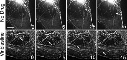
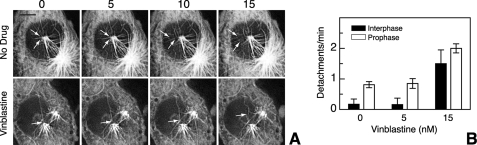
Drugs that target microtubules are thought to inhibit cell division and cell migration by suppressing dynamic instability, a "search and capture" behavior that allows microtubules to probe their environment. Here, we report that subtoxic drug concentrations are sufficient to inhibit plus-end microtubule dynamic instability and cell migration without affecting cell division or microtubule assembly. The higher drug concentrations needed to inhibit cell division act through a novel mechanism that generates microtubule fragments by stimulating microtubule minus-end detachment from their organizing centers. The frequency of microtubule detachment in untreated cells increases at prophase suggesting that it is a regulated cellular process important for spindle assembly and function. We conclude that drugs produce differential dose-dependent effects at microtubule plus and minus-ends to inhibit different microtubule-mediated functions.
Figures
Similar articles
-
Detection and quantification of microtubule detachment from centrosomes and spindle poles.Methods Cell Biol. 2013;115:49-62. doi: 10.1016/B978-0-12-407757-7.00004-9. Methods Cell Biol. 2013. PMID: 23973065
-
Megakaryocyte lineage-specific class VI β-tubulin suppresses microtubule dynamics, fragments microtubules, and blocks cell division.Cytoskeleton (Hoboken). 2011 Mar;68(3):175-87. doi: 10.1002/cm.20503. Cytoskeleton (Hoboken). 2011. PMID: 21309084 Free PMC article.
-
Paclitaxel-dependent cell lines reveal a novel drug activity.Mol Cancer Ther. 2010 Nov;9(11):2914-23. doi: 10.1158/1535-7163.MCT-10-0552. Epub 2010 Oct 26. Mol Cancer Ther. 2010. PMID: 20978163 Free PMC article.
-
Control of microtubule organization and dynamics: two ends in the limelight.Nat Rev Mol Cell Biol. 2015 Dec;16(12):711-26. doi: 10.1038/nrm4084. Epub 2015 Nov 12. Nat Rev Mol Cell Biol. 2015. PMID: 26562752 Review.
-
Interplay between microtubule dynamics and intracellular organization.Int J Biochem Cell Biol. 2012 Feb;44(2):266-74. doi: 10.1016/j.biocel.2011.11.009. Epub 2011 Nov 17. Int J Biochem Cell Biol. 2012. PMID: 22108200 Review.
Cited by
-
Moving toward a higher efficiency of microcell-mediated chromosome transfer.Mol Ther Methods Clin Dev. 2016 Jun 22;3:16043. doi: 10.1038/mtm.2016.43. eCollection 2016. Mol Ther Methods Clin Dev. 2016. PMID: 27382603 Free PMC article.
-
Non-linear Dose Response of Lymphocyte Cell Lines to Microtubule Inhibitors.Front Pharmacol. 2019 Apr 24;10:436. doi: 10.3389/fphar.2019.00436. eCollection 2019. Front Pharmacol. 2019. PMID: 31068822 Free PMC article.
-
Casein kinase I delta controls centrosome positioning during T cell activation.J Cell Biol. 2011 Nov 28;195(5):781-97. doi: 10.1083/jcb.201106025. J Cell Biol. 2011. PMID: 22123863 Free PMC article.
-
HYS-32-Induced Microtubule Catastrophes in Rat Astrocytes Involves the PI3K-GSK3beta Signaling Pathway.PLoS One. 2015 May 4;10(5):e0126217. doi: 10.1371/journal.pone.0126217. eCollection 2015. PLoS One. 2015. PMID: 25938237 Free PMC article.
-
Drug conjugates for the treatment of lung cancer: from drug discovery to clinical practice.Exp Hematol Oncol. 2024 Mar 1;13(1):26. doi: 10.1186/s40164-024-00493-8. Exp Hematol Oncol. 2024. PMID: 38429828 Free PMC article. Review.
References
-
- Mitchison T., Kirschner M. W. (1984) Nature 312, 237–242 - PubMed
-
- Small J. V., Geiger B., Kaverina I., Bershadsky A. (2002) Nat. Rev. Mol. Cell Biol. 3, 957–964 - PubMed
-
- Jordan M. A., Wilson L. (2004) Nature Reviews 4, 253–265 - PubMed
-
- Pourroy B., Honoré S., Pasquier E., Bourgarel-Rey V., Kruczynski A., Briand C., Braguer D. (2006) Cancer Res. 66, 3256–3263 - PubMed
Publication types
MeSH terms
Substances
Grants and funding
LinkOut - more resources
Full Text Sources
