. 2010 Oct 15;285(42):32494-503.
doi: 10.1074/jbc.M110.142430. Epub 2010 Aug 9.

A lethal de novo mutation in the middle domain of the dynamin-related GTPase Drp1 impairs higher order assembly and mitochondrial division

Affiliations

A lethal de novo mutation in the middle domain of the dynamin-related GTPase Drp1 impairs higher order assembly and mitochondrial division

Chuang-Rung Chang et al. J Biol Chem. .

Abstract

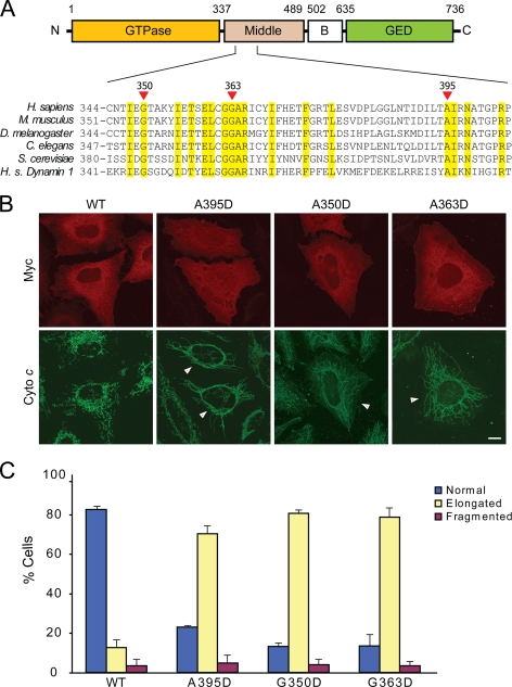
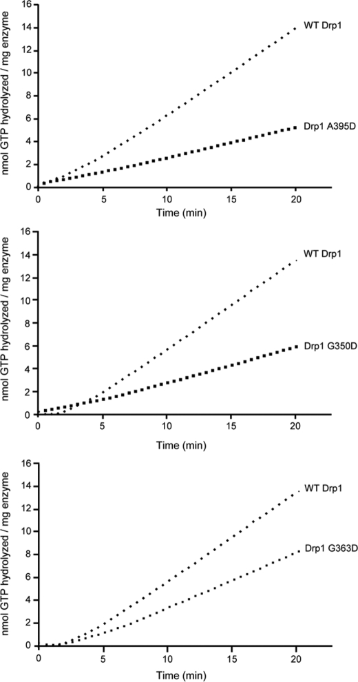
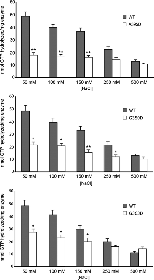
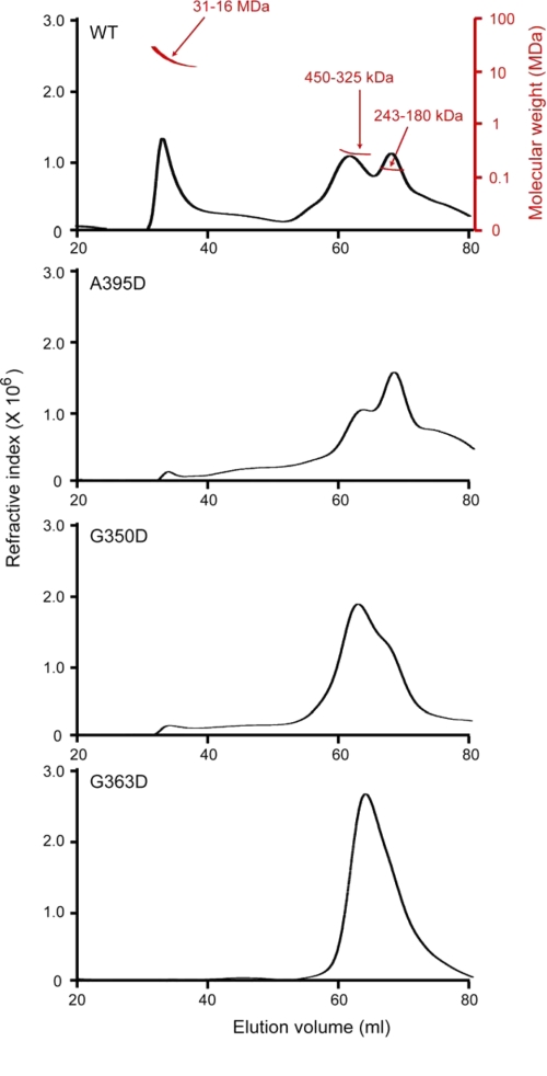
Mitochondria dynamically fuse and divide within cells, and the proper balance of fusion and fission is necessary for normal mitochondrial function, morphology, and distribution. Drp1 is a dynamin-related GTPase required for mitochondrial fission in mammalian cells. It harbors four distinct domains: GTP-binding, middle, insert B, and GTPase effector. A lethal mutation (A395D) within the Drp1 middle domain was reported in a neonate with microcephaly, abnormal brain development, optic atrophy, and lactic acidemia (Waterham, H. R., Koster, J., van Roermund, C. W., Mooyer, P. A., Wanders, R. J., and Leonard, J. V. (2007) N. Engl. J. Med. 356, 1736-1741). Mitochondria within patient-derived fibroblasts were markedly elongated, but the molecular mechanisms underlying these findings were not demonstrated. Because the middle domain is particularly important for the self-assembly of some dynamin superfamily proteins, we tested the hypothesis that this A395D mutation, and two other middle domain mutations (G350D, G363D) were important for Drp1 tetramerization, higher order assembly, and function. Although tetramerization appeared largely intact, each of these mutations compromised higher order assembly and assembly-dependent stimulation of Drp1 GTPase activity. Moreover, mutant Drp1 proteins exhibited impaired localization to mitochondria, indicating that this higher order assembly is important for mitochondrial recruitment, retention, or both. Overexpression of these middle domain mutants markedly inhibited mitochondrial division in cells. Thus, the Drp1 A395D lethal defect likely resulted in impaired higher order assembly of Drp1 at mitochondria, leading to decreased fission, elongated mitochondria, and altered cellular distribution of mitochondria.


Figures

FIGURE 1.
FIGURE 1.
Drp1 middle domain mutations result in elongated mitochondrial morphology. A, top, shown is the schematic of human Drp1 structure (splice variant 1) with boundary amino acids shown for domains. Bottom, shown is phylogenetic sequence alignment of the depicted middle domain region of Drp1 orthologs in the indicated species. Homo sapiens (H. s.) dynamin-1 is included at the bottom. Yellow shading identifies residues identical in all proteins shown. Red arrows identify conserved residues investigated in this study (numbering is shown for human Drp1). B, HeLa cells were transfected with WT or the indicated Drp1 mutants and visualized by confocal immunofluorescence microscopy. Transfected cells are identified by arrows in the lower panels. C, mitochondrial morphology was quantified as described under “Experimental Procedures.” Bar, 10 μm.
FIGURE 2.
FIGURE 2.
Middle domain mutations decrease Drp1 recruitment/retention on mitochondria. A, expression of the Myc-tagged wild-type (WT) and mutant forms of Drp1 was equal. Actin levels were monitored as a loading control. B, HeLa cells transfected with WT Myc-Drp1, or else the indicated Drp1 mutants were either left untreated or treated with staurosporine (STS) in the presence of the broad caspase inhibitor benzyloxycarbonyl-VAD-fluoromethyl ketone. Levels of Drp1 in the mitochondrial fraction were assessed by immunoblotting, with levels of the mitochondrial protein HSP60 monitored as a loading control.
FIGURE 3.
FIGURE 3.
Drp1 middle domain mutants do not affect Drp1 self-association. A, co-immunoprecipitation of Myc- and HA-tagged Drp1 proteins is shown. HeLa cells expressing the indicated WT or mutant Drp1 proteins were immunoprecipitated with anti-HA antibodies (αHA IP) and immunoblotted with anti-Myc antibodies. Input represents 20% of the starting lysate. The IgG heavy chain from the precipitating antibodies is noted with an asterisk (*). B, cytosolic extracts from cells overexpressing wild-type or mutant Myc-Drp1 were cross-linked with the indicated concentrations of bis(sulfosuccinimidyl) suberate (BS3), resolved by SDS-PAGE and immunoblotted with anti-Myc antibodies. Migrations of molecular mass standards are indicated at the left. A prominent cross-linked product at ∼280 kDa is indicated with an arrowhead in B.
FIGURE 4.
FIGURE 4.
Middle domain mutations disrupt intra- and intermolecular interactions of full-length Drp1. Yeast two-hybrid assays using the HIS3 reporter (sequential 10-fold yeast dilutions are shown) with the indicated bait and prey constructs are shown for intramolecular (A) and intermolecular (B) Drp1 interactions. Drp1 mutations across the top are present in constructs identified as mut. Arrows, arrowheads, and asterisks (*) are referenced under “Results.”
FIGURE 5.
FIGURE 5.
Middle domain mutations disrupt intermolecular interactions mediated by Drp1 middle domains. A and B, yeast two-hybrid assays using the HIS3 reporter (sequential 10-fold yeast dilutions are shown) with the indicated bait and prey constructs are shown. Arrows and arrowheads are referenced under “Results.”
FIGURE 6.
FIGURE 6.
Drp1 middle domain mutations decrease assembly-dependent GTP hydrolysis. GTP hydrolysis by 2.5 μm CBP fusions of WT Drp1, and the indicated Drp1 mutants were measured in a coupled, substrate-regenerative GTPase assay as described under “Experimental Procedures.” Each trace shown is representative of experiments comprising at least three independent preparations of Drp1 performed in triplicate.
FIGURE 7.
FIGURE 7.
Drp1 middle domain mutations alter salt-dependent GTPase activities. The total amount of GTP hydrolyzed by 2.5 μm Drp1 or Drp1 mutants was assayed as a function of [NaCl] for 40 min. Data shown are averages of three independent preparations of CBP fusions of WT Drp1 and Drp1 A395D (A), Drp1 G350D (B), and Drp1 G363D (C) measured in triplicate ± S.E. **, p < 0.005; *, p < 0.05.
FIGURE 8.
FIGURE 8.
Drp1 middle domain mutants are higher order assembly-deficient dimers and tetramers. CBP-Drp1 WT and mutant fusion proteins were analyzed in 250 mm NaCl by SEC-MALS using a Superose 6 10/30 HR column as described under “Experimental Procedures.” Refractive index is plotted as a function of elution volume (ml). A molecular weight scale based on MALS analysis for eluted protein is shown for WT Drp1 (top panel; red) and is similar for each of the mutant proteins. WT Drp1 eluted primarily as 3 peaks, one that corresponds to a molecular mass range of 31–16 MDa that eluted in the void volume and two peaks that corresponded to molecular mass ranges of 420–325 and 243–180 kDa. This is consistent with an equilibrium between dimer-tetramer and higher order species given that the expected molecular mass for Drp1 dimer is ∼180 kDa and tetramer is ∼360 kDa. Mutant proteins predominantly comprise dimer and tetramer species, with little to no assembled protein in the column void volume, consistent with a defect in higher order assembly.

References

    1. Li Z., Okamoto K., Hayashi Y., Sheng M. (2004) Cell 119, 873–887 - PubMed
    1. Arnoult D., Rismanchi N., Grodet A., Roberts R. G., Seeburg D. P., Estaquier J., Sheng M., Blackstone C. (2005) Curr. Biol. 15, 2112–2118 - PubMed
    1. Campello S., Lacalle R. A., Bettella M., Mañes S., Scorrano L., Viola A. (2006) J. Exp. Med. 203, 2879–2886 - PMC - PubMed
    1. Chan D. C. (2006) Cell 125, 1241–1252 - PubMed
    1. McBride H. M., Neuspiel M., Wasiak S. (2006) Curr. Biol. 16, R551–R560 - PubMed

Publication types

MeSH terms

LinkOut - more resources