. 2010 Oct 15;285(42):32576-85.
doi: 10.1074/jbc.M110.153817. Epub 2010 Aug 9.

Rod/Zw10 complex is required for PIASy-dependent centromeric SUMOylation

Affiliations

Rod/Zw10 complex is required for PIASy-dependent centromeric SUMOylation

Hyunju Ryu et al. J Biol Chem. .

Abstract

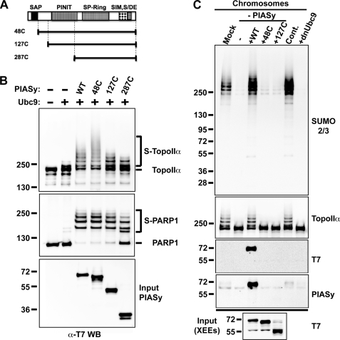
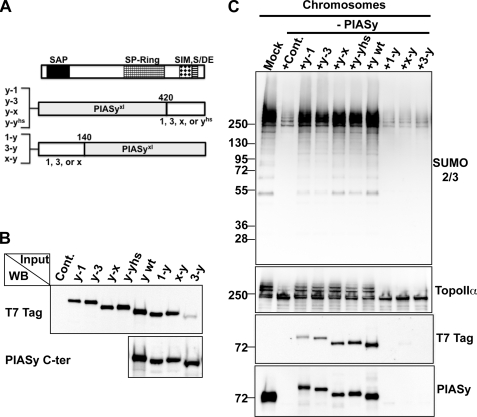
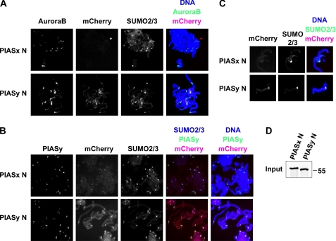
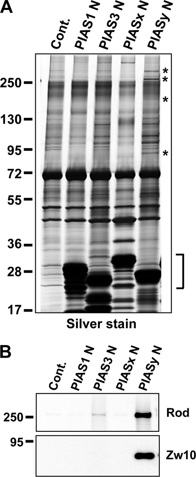
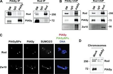
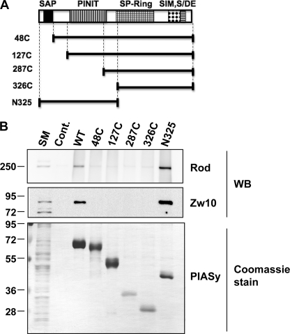
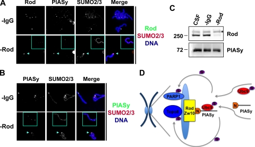
SUMO conjugation of cellular proteins is essential for proper progression of mitosis. PIASy, a SUMO E3 ligase, is required for mitotic SUMOylation of chromosomal proteins, yet the regulatory mechanism behind the PIASy-dependent SUMOylation during mitosis has not been determined. Using a series of truncated PIASy proteins, we have found that the N terminus of PIASy is not required for SUMO modification in vitro but is essential for mitotic SUMOylation in Xenopus egg extracts. We demonstrate that swapping the N terminus of PIASy protein with the corresponding region of other PIAS family members abolishes chromosomal binding and mitotic SUMOylation. We further show that the N-terminal domain of PIASy is sufficient for centromeric localization. We identified that the N-terminal domain of PIASy interacts with the Rod/Zw10 complex, and immunofluorescence further reveals that PIASy colocalizes with Rod/Zw10 in the centromeric region. We show that the Rod/Zw10 complex interacts with the first 47 residues of PIASy which were particularly important for mitotic SUMOylation. Finally, we show that depletion of Rod compromises the centromeric localization of PIASy and SUMO2/3 in mitosis. Together, we demonstrate a fundamental mechanism of PIASy to localize in the centromeric region of chromosome to execute centromeric SUMOylation during mitosis.


Figures

FIGURE 1.
FIGURE 1.
N terminus of PIASy is dispensable in vitro but essential for mitotic SUMOylation in Xenopus egg extract assays. A, schematic diagram of PIASy truncations used in experiments in B and C. N-terminal deletions of PIASy are drawn to approximate scale and compared with full-length PIASy shown on the top. All truncation constructs and full-length PIASy contain a T7 tag at the N terminus. Conserved domains among PIAS family are indicated as follows: SAP in solid black, PINIT in vertical lines, SP-Ring in grid, SIM in diamond, and S/DE motif in horizontal lines. B, in vitro SUMOylation assay using N-terminal-truncated PIASy series. All constructs were expressed and purified as described under “Experimental Procedures.” Either TopoIIα or PARP1 was incubated with PIASy and Ubc9 as indicated in the presence of E1 and ATP for 1 h at 25 °C, and samples were analyzed by Western blotting with anti-T7 tag antibody, which detects recombinant TopoIIα and PARP1. SUMOylated and unmodified forms of proteins are specified by bracket and bar, respectively. The PIASy input is shown in the bottom panel. C, PIASy immunodepletion and addback experiments in Xenopus egg extracts (XEE). CSF-XEE was immunodepleted using IgG (Mock) or anti-PIASy antibody. Purified PIASy truncation proteins were added to the depleted XEE and 5,000 sperm chromatin/μl was incubated in the reactions at 25 °C for ∼1 h. The reactions without (Cont.) and with dnUbc9 were also prepared for positive and negative control of chromosomal SUMOylation. Isolated chromosome fractions were resolved on SDS-PAGE gel and analyzed by Western blot for indicated proteins. The input of PIASy proteins were analyzed in the bottom panel.
FIGURE 2.
FIGURE 2.
Comparison of function of PIAS N-terminal domains in the XEE assay. A, schematic diagram of PIAS chimera proteins. The chimeric PIAS proteins with T7 tag at the N terminus were obtained by swapping conserved domains between PIAS family members as described under “Experimental Procedures.” The diagram indicates the positions of swapped domain between PIAS family proteins. 1, PIAS1; 3, PIAS3; x, PIASxα; yhs, human PIASy; and y, Xenopus PIASy. B, expression of the chimeric PIAS proteins in reticulocyte lysates. The chimera proteins constructed in A were expressed in rabbit reticulocyte lysates, and the expression level was analyzed by Western blot for T7 tag or anti-C terminus of PIASy antibody. The reticulocyte lysate containing the empty vector was prepared for the negative control (Cont.). C, immunodepletion and chimeric PIAS addback experiments in XEEs. CSF-XEE was immunodepleted by IgG (Mock) or anti-PIASy antibody. Reticulocyte lysates shown in B were added to the PIASy-depleted CSF extracts, and the extracts were incubated in the presence of 5,000 sperm chromatin/μl for ∼1 h at 25 °C. Isolated chromosomes were analyzed by Western blot for indicated proteins.
FIGURE 3.
FIGURE 3.
N-terminal region of PIASy locates to centromere independent of its remaining residues. A and B, localization of mCherry-tagged N-terminal peptides of PIASx and PIASy. Replicated chromosomes were obtained by incubating 500 sperm nuclei/μl in interphase extract followed by re-entry into mitosis by CSF-XEE. N-terminal peptides of PIASx (PIASx N) or PIASy (PIASy N) that have mCherry fused at the C terminus were added before re-entry into mitosis. After re-induction into mitosis by fresh CSF-XEE, chromosomes were spun down on coverslips by centrifugation. Samples were immunostained for indicated proteins. C, centromeric localization of mCherry-fused N-terminal peptide of PIASy. Single sister chromatids are enlarged for the comparison of localization between PIASx N and PIASy N. D, input of PIASx N and PIASy N. The amount of PIASx N and PIASy N input was analyzed by Western blot with S-protein HRP (EMD Bioscience), which detects the S tag at the N terminus on both PIASx N and PIASy N peptides.
FIGURE 4.
FIGURE 4.
Identification of N-terminal-binding proteins of PIASy. A, N terminus of PIASy has distinctive binding proteins. S-tagged N-terminal domains of PIAS family proteins were bound to S-agarose beads. Either S-agarose beads (Cont.) or PIAS N-terminal protein-bound bead preparations were incubated in CSF XEE. Precipitated proteins together with beads were resolved by SDS-PAGE followed by silver staining. Specific binding proteins for PIASy N-terminal domain are indicated with an asterisk. S-tagged PIAS N-terminal peptides used as bait are indicated with a bracket. B, confirmation of Rod and Zw10 binding to PIASy N terminus. Pull-down samples as in A were analyzed by Western blot for Rod and Zw10. The antibody against Rod, prepared as described under “Experimental Procedures,” or commercially available anti-Zw10 antibody (AbCam) were used to detect them in pull-down samples.
FIGURE 5.
FIGURE 5.
PIASy interacts with Rod/Zw10 complex on mitotic chromosomes. A, interaction of Rod and Zw10 proteins with endogenous PIASy in CSF-XEE. Left, IgG or anti-PIASy antibody cross-linked protein A beads were incubated with CSF-XEE. Immunoprecipitated proteins were resolved in SDS-PAGE and analyzed by Western blot for indicated proteins. Right, IgG or anti-Rod antibody cross-linked protein A beads were incubated in CSF-XEE in the same manner. The precipitants were resolved by SDS-PAGE followed by Western blot. SM stands for starting material. The band labeled with asterisk is a nonspecific protein cross-reacting with anti-Rod antibody in CSF-XEE. IP, immunoprecipitation. B, interaction of PIASy and Rod/Zw10 complex on chromosomes. Mitotic chromosomes prepared in CSF-XEE were subjected to chromatin immunoprecipitation as described under “Experimental Procedures,” and precipitated samples were analyzed as in A. C, localization of endogenous PIASy and binding proteins, Rod and Zw10. Replicated mitotic chromosome samples were prepared as in Fig. 3 followed by immunostaining for the indicated proteins. PIASyBPs, PIASy-binding proteins. D, depletion of PIASy does not eliminate the interaction of Rod/Zw10 complex to mitotic chromosomes. Mock-depleted by IgG (Mock) or PIASy-depleted CSF-XEE (-PIASy) were incubated in the presence of 5,000 sperm nuclei/μl for 1 h at 25 °C. Isolated chromosomes were resolved by SDS-PAGE and analyzed by Western blot. The antibody cross-reacting band is indicated with asterisk.
FIGURE 6.
FIGURE 6.
Rod/Zw10 complex interacts with the first 47 residues of PIASy protein. A, schematic diagram of PIASy and its truncations used in B. PIASy and its truncations contain T7 tag at the N terminus. B, further identification of Rod/Zw10-interacting area on PIASy protein. Purified T7-tagged PIASy truncation proteins as well as full-length (WT) were bound to T7 antibody-conjugated agarose beads. Protein-bound beads or beads alone (Cont.) were incubated with CSF-XEE. The precipitated proteins together with the T7-antibody beads were resolved by SDS-PAGE and analyzed by Western blot for the indicated proteins or Coomassie Blue staining. SM, starting material.
FIGURE 7.
FIGURE 7.
Rod/Zw10 complex is required for localization of PIASy on the centromeric region as well as SUMO2/3 during mitosis. A and B, immunodepletion of Rod causes mislocalization of PIASy on mitotic chromosomes. A, CSF-XEE was immunodepleted for Rod and released into interphase by the addition of CaCl2. Sperm chromatin was incubated with the interphase extracts, and sister chromatids were obtained from the replicated chromatin by the addition of Rod-depleted CSF-XEE (−Rod). CSF-XEE was also mock-depleted using nonspecific IgG and processed as above (−IgG). Isolated chromosome samples were subjected to immunostaining for proteins as indicated. Affinity-purified anti-PIASy chicken IgY and anti-SUMO2/3 guinea pig IgG were used to visualize PIASy and SUMO2/3, respectively. B, samples in A were immunostained with affinity purified anti-PIASy antibody obtained from rabbit. The chromosome lacking Rod protein is shown in squares. The chromosome that still contains Rod in Rod-depleted samples is indicated with arrows. The centromere-specific localization of both PIASy and SUMO modification was compromised if Rod was completely eliminated. C, confirmation of Rod depletion. CSF-XEE before and after immunodepletion with anti-IgG or Rod antibody were analyzed for the indicated proteins. Asterisk shows background signals from antibody cross-reacting in CSF extracts. D, model for molecular mechanism of PIASy-dependent centromeric SUMOylation during mitosis. PIASy interacts with the Rod/Zw10 complex at the kinetochore through its N-terminal region (designated as N). Consequently, Ubc9-SUMO2/3 adduct is recruited onto centromeric regions via binding to PIASy and facilitates SUMO2/3 modification of substrates such as TopoIIα and PARP1 in a spatially regulated manner.

Similar articles

Cited by

References

    1. Johnson E. S. (2004) Annu. Rev. Biochem. 73, 355–382 - PubMed
    1. Bossis G., Melchior F. (2006) Cell Div. 1, 13. - PMC - PubMed
    1. Dasso M. (2008) Cell Div. 3, 5. - PMC - PubMed
    1. Hay R. T. (2005) Mol. Cell 18, 1–12 - PubMed
    1. Bernier-Villamor V., Sampson D. A., Matunis M. J., Lima C. D. (2002) Cell 108, 345–356 - PubMed

Publication types

MeSH terms

Substances

LinkOut - more resources