

Specific and nonspecific B-cell function in the small intestines of patients with Whipple's disease
- PMID: 20696822
- PMCID: PMC2976328
- DOI: 10.1128/IAI.00705-10
Specific and nonspecific B-cell function in the small intestines of patients with Whipple's disease
Abstract
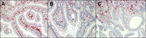
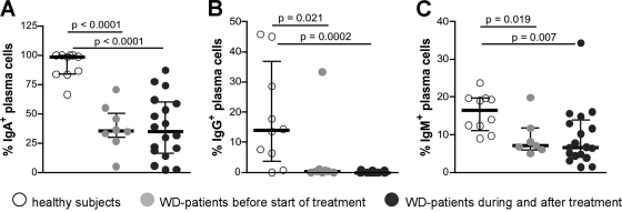
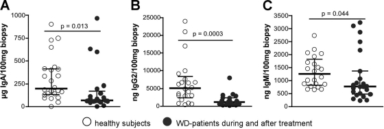
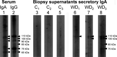
Whipple's disease is a chronic multisystemic infection caused by Tropheryma whipplei that is characterized by arthritis, weight loss, and diarrhea. The immunological defects in the duodenal mucosa, the site of major replication of the agent underlying the pathogenesis of Whipple's disease, are poorly understood. Mucosal immunoglobulins are essential for the defense against intestinal pathogens; therefore, we analyzed the B-cell response in duodenal specimens and sera of Whipple's disease patients. Whereas systemic immunoglobulin production was affected only marginally, duodenal biopsy specimens of Whipple's disease patients contained reduced numbers of immunoglobulin-positive plasma cells and secreted less immunoglobulin compared to healthy controls but showed a weak secretory IgA response toward T. whipplei. This T. whipplei-specific intestinal immune response was not observed in controls. Thus, we were able to demonstrate that general mucosal immunoglobulin production in Whipple's disease patients is impaired. However, this deficiency does not completely abolish T. whipplei-specific secretory IgA production that nonetheless does not protect from chronic infection.
Figures
Similar articles
-
Changing paradigms in Whipple's disease and infection with Tropheryma whipplei.Eur J Clin Microbiol Infect Dis. 2011 Oct;30(10):1151-8. doi: 10.1007/s10096-011-1209-y. Epub 2011 Apr 2. Eur J Clin Microbiol Infect Dis. 2011. PMID: 21461659 Review.
-
Peripheral T-Cell Reactivity to Heat Shock Protein 70 and Its Cofactor GrpE from Tropheryma whipplei Is Reduced in Patients with Classical Whipple's Disease.Infect Immun. 2017 Jul 19;85(8):e00363-17. doi: 10.1128/IAI.00363-17. Print 2017 Aug. Infect Immun. 2017. PMID: 28559404 Free PMC article.
-
Impaired immune functions of monocytes and macrophages in Whipple's disease.Gastroenterology. 2010 Jan;138(1):210-20. doi: 10.1053/j.gastro.2009.07.066. Epub 2009 Aug 5. Gastroenterology. 2010. PMID: 19664628
-
Reduced peripheral and mucosal Tropheryma whipplei-specific Th1 response in patients with Whipple's disease.J Immunol. 2006 Aug 1;177(3):2015-22. doi: 10.4049/jimmunol.177.3.2015. J Immunol. 2006. PMID: 16849516
-
[First centenary of Whipple's disease].Rev Alerg Mex. 2009 May-Jun;56(3):92-8. Rev Alerg Mex. 2009. PMID: 19623786 Review. Spanish.
Cited by
-
Changing paradigms in Whipple's disease and infection with Tropheryma whipplei.Eur J Clin Microbiol Infect Dis. 2011 Oct;30(10):1151-8. doi: 10.1007/s10096-011-1209-y. Epub 2011 Apr 2. Eur J Clin Microbiol Infect Dis. 2011. PMID: 21461659 Review.
-
Role of dendritic cells in the pathogenesis of Whipple's disease.Infect Immun. 2015 Feb;83(2):482-91. doi: 10.1128/IAI.02463-14. Epub 2014 Nov 10. Infect Immun. 2015. PMID: 25385798 Free PMC article.
-
Fever and Increased Gastrointestinal Uptake on Positron Emission Tomography after Anti-Tumour Necrosis Factor Therapy: A Case Report of Whipple's Disease.Case Rep Gastroenterol. 2024 Apr 20;18(1):221-230. doi: 10.1159/000538462. eCollection 2024 Jan-Dec. Case Rep Gastroenterol. 2024. PMID: 38645407 Free PMC article.
-
Architectural and functional alterations of the small intestinal mucosa in classical Whipple's disease.Mucosal Immunol. 2017 Nov;10(6):1542-1552. doi: 10.1038/mi.2017.6. Epub 2017 Feb 8. Mucosal Immunol. 2017. PMID: 28176790
-
Peripheral T-Cell Reactivity to Heat Shock Protein 70 and Its Cofactor GrpE from Tropheryma whipplei Is Reduced in Patients with Classical Whipple's Disease.Infect Immun. 2017 Jul 19;85(8):e00363-17. doi: 10.1128/IAI.00363-17. Print 2017 Aug. Infect Immun. 2017. PMID: 28559404 Free PMC article.
References
-
- Bonhomme, C. J., P. Renesto, B. Desnues, E. Ghigo, H. Lepidi, P. Fourquet, F. Fenollar, B. Henrissat, J. L. Mege, and D. Raoult. 2009. Tropheryma whipplei glycosylation in the pathophysiologic profile of Whipple's disease. J. Infect. Dis. 199:1043-1052. - PubMed
-
- Bonhomme, C. J., P. Renesto, S. Nandi, A. M. Lynn, and D. Raoult. 2008. Serological microarray for a paradoxical diagnostic of Whipple's disease. Eur. J. Clin. Microbiol. Infect. Dis. 27:959-968. - PubMed
-
- Desnues, B., H. Lepidi, D. Raoult, and J. L. Mege. 2005. Whipple disease: intestinal infiltrating cells exhibit a transcriptional pattern of M2/alternatively activated macrophages. J. Infect. Dis. 192:1642-1646. - PubMed
-
- Ectors, N., K. Geboes, R. De Vos, H. Heidbuchel, P. Rutgeerts, V. Desmet, and G. Vantrappen. 1992. Whipple's disease: a histological, immunocytochemical, and electron microscopic study of the immune response in the small intestinal mucosa. Histopathology 21:1-12. - PubMed
-
- Fenollar, F., B. Amphoux, and D. Raoult. 2009. A paradoxical Tropheryma whipplei Western blot differentiates patients with Whipple disease from asymptomatic carriers. Clin. Infect. Dis. 49:717-723. - PubMed
Publication types
MeSH terms
Substances
LinkOut - more resources
Full Text Sources
Miscellaneous
