. 2010 Oct;78(10):4166-75.
doi: 10.1128/IAI.00711-10. Epub 2010 Aug 9.

Enterococcal biofilm formation and virulence in an optimized murine model of foreign body-associated urinary tract infections

Affiliations

Enterococcal biofilm formation and virulence in an optimized murine model of foreign body-associated urinary tract infections

Pascale S Guiton et al. Infect Immun. 2010 Oct.

Abstract

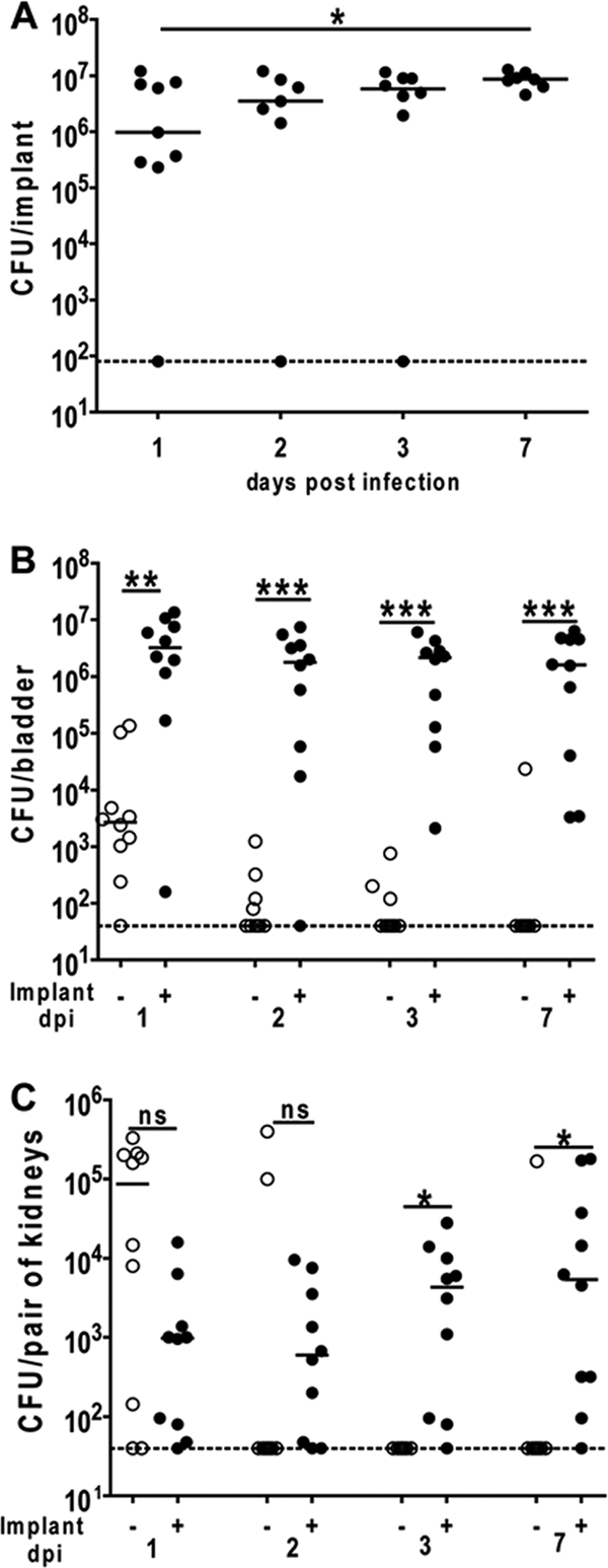
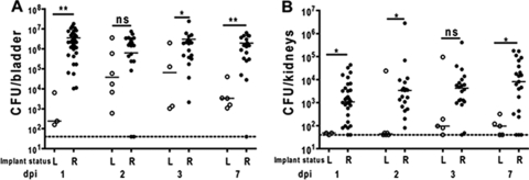
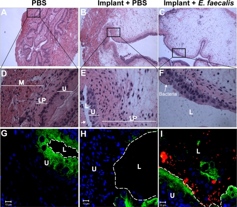
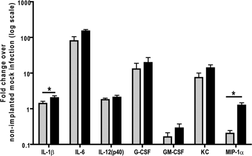
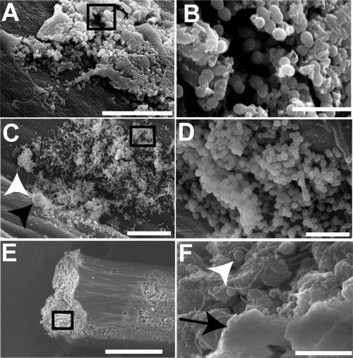
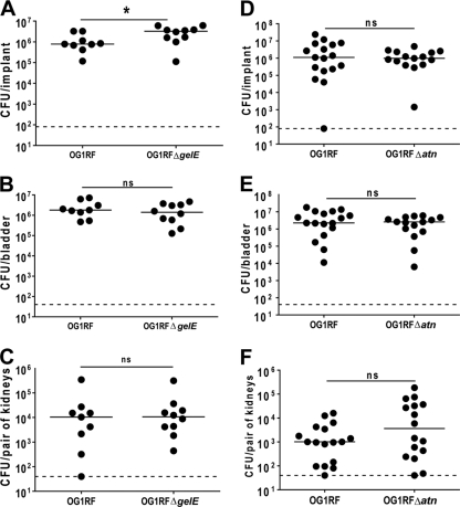
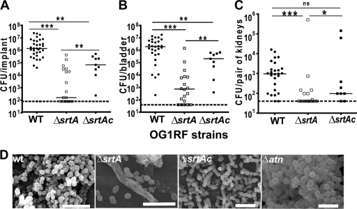
Catheter-associated urinary tract infections (CAUTIs) constitute the majority of nosocomial UTIs and pose significant clinical challenges. Enterococcal species are among the predominant causative agents of CAUTIs. However, very little is known about the pathophysiology of Enterococcus-mediated UTIs. We optimized a murine model of foreign body-associated UTI in order to mimic conditions of indwelling catheters in patients. In this model, the presence of a foreign body elicits major histological changes and induces the expression of several proinflammatory cytokines in the bladder. In addition, in contrast to naïve mice, infection of catheter-implanted mice with Enterococcus faecalis induced the specific expression of interleukin 1β (IL-1β) and macrophage inflammatory protein 1α (MIP-1α) in the bladder. These responses resulted in a favorable niche for the development of persistent E. faecalis infections in the murine bladders and kidneys. Furthermore, biofilm formation on the catheter implant in vivo correlated with persistent infections. However, the enterococcal autolytic factors GelE and Atn (also known as AtlA), which are important in biofilm formation in vitro, are dispensable in vivo. In contrast, the housekeeping sortase A (SrtA) is critical for biofilm formation and virulence in CAUTIs. Overall, this murine model represents a significant advance in the understanding of CAUTIs and underscores the importance of urinary catheterization during E. faecalis uropathogenesis. This model is also a valuable tool for the identification of virulence determinants that can serve as potential antimicrobial targets for the treatment of enterococcal infections.


Figures

FIG. 1.
FIG. 1.
E. faecalis establishes persistent colonization of silicone implants, bladders, and kidneys in a murine model of CAUTI. (A) Bacterial titers in logarithmic scale (log scale) from silicone implants retrieved from female C57BL/6Ncr mice infected with OG1RF for 24, 48, and 72 hpi (1, 2, and 3 dpi) and 7 dpi. (B and C) Graphs represent bacterial titers from homogenized bladders (B) and kidneys (C) from animals without implants (−) (○) or animals with implants (+) (•) infected with strain OG1RF for 1, 2, 3, or 7 dpi. In panels A to C, the horizontal broken line represents the limit of detection for viable bacteria. Each symbol represents the value for an individual mouse from two independent experiments with five mice for each condition. The horizontal bar represents the median value for each group of mice. Values that are significantly different by the Mann-Whitney U test are indicated as follows: *, P < 0.05; **, P < 0.005; ***, P < 0.0005. Values that are not significantly different (ns) by the Mann-Whitney U test are indicated.
FIG. 2.
FIG. 2.
Implant loss from murine bladders correlates with less severe infection of the bladder and kidney. Graphs represent bacterial titers in log scale recovered from the bladders (A) and kidneys (B) of animals that have lost the implants (L) (○) or retained the implants (R) (•) at the indicated time of sacrifice. The experiment was done at least three times with five mice in each experiment. The horizontal broken line depicts the limit of detection. The horizontal bar represents the median value for each group of mice. Values that are significantly different by the Mann-Whitney U test are indicated as follows: *, P < 0.05; **, P < 0.005; ***, P < 0.0005. Values that are not significantly different (P > 0.05) (ns) by the Mann-Whitney U test are indicated.
FIG. 3.
FIG. 3.
Implantation-mediated histological changes are associated with enterococcal colonization of the murine bladder. (A to F) H&E staining of bladder sections derived from animals without implants inoculated with PBS (A and D), animals with implants inoculated with PBS (B and E), or animals with implants infected with E. faecalis OG1RF (C and F) 24 hpi observed with a light microscope at magnifications of ×40 (A to C) and ×63 (D to F). Areas in black boxes on panels A, B, and C are magnified in panels D, E, and F, respectively. Severe edema, hyperplasia, urothelial sloughing, and immune cell infiltration characterize bladders of implanted animals with or without bacterial challenge. The white arrow in panel E indicates damage to the uroepithelium. Abbreviations: U, uroepithelium; LP, lamina propria; L, lumen; M, muscularis. (G to I) Representative laser scanning confocal microscopic (CLSM) images of bladder sections from PBS-treated nonimplanted, PBS-treated implanted, and OG1RF-infected implanted mice 24 hpi immunolabeled with goat anti-mouse uroplakin III (green), rabbit anti-Lancefield group D antigen (red), and counterstained with TOPRO-3 nuclear dye (blue) reveal alterations to the uroepithelium of implanted bladders and the presence of bacteria in the bladder lumen and on the uroepithelial surface. The white broken line separates the lumen (L) from the uroepithelium surface (U). Bars = 10 μm.
FIG. 4.
FIG. 4.
Implantation and E. faecalis infection elicit the production of specific cytokines in the bladder. Graphs represent cytokine expression in the whole bladder of animals with implants inoculated with PBS or E. faecalis OG1RF at 24 hpi. Cytokines reported are expressed with at least twofold difference relative to mock-infected animals with no implants. The presence of implants induces the specific expression of IL-6, G-CSF, and KC and causes the downregulation of GM-CSF and MIP-1α. In contrast, E. faecalis elicits the specific expression of IL-1β, IL-12(p40), and MIP-1α at 24 hpi. Data represent a representative experiment performed four times with five mice for each condition in the experiment. Values are shown as the means plus standard errors of the means (error bars). Values that are significantly different (P < 0.05 by the Mann-Whitney U test) are indicated (*).
FIG. 5.
FIG. 5.
E. faecalis produces biofilms on the surfaces of silicone implants in vivo. Representative scanning electron micrographs of silicone implants retrieved from murine bladders infected with E. faecalis OG1RF at 72 hpi showing bacterial biofilms on the outer surface (A and B) and filling the lumen (C and F) of the silicone implant. Bacteria (white arrowhead) are often associated with host cells, indicated by the black arrowhead in panel C. Both bacteria and host immune cells can also be found embedded in a thick extracellular material, indicated by the black arrow, in the lumen of the silicone implant 72 hpi (E and F). Areas in black boxes on panels A, C, and E are magnified in panels B, D, and F, respectively. Bars, 20 μm (A and C), 5 μm (B, D, and F), and 100 μm (E).
FIG. 6.
FIG. 6.
Autolytic factors are not required for E. faecalis virulence in the urinary tract. (A to F) Graphs show the numbers of CFU of E. faecalis OG1RF and isogenic in-frame gelE- and atn-deficient mutants recovered from retrieved implants (A and D), bladders (B and E), and kidneys (C and F) from animals 24 hpi following infection with either strain. Horizontal dashed lines represent the limits of detection for viable bacteria. Each symbol represents the value for an individual mouse, and each experiment was done twice with four or five mice for each strain. Only data from animals with implants at the time of sacrifice are represented. The horizontal bars indicate the median values for groups. Values that are statistically significantly different (P < 0.05) by the Mann-Whitney U test are indicated (*). Values that are not statistically significantly different (P > 0.05) by the Mann-Whitney U test are indicated (ns).
FIG. 7.
FIG. 7.
Sortase A is critical for implant colonization and virulence in the urinary tract. (A to C) Graphs show bacterial titers from implants (A), homogenized bladders (B), and kidneys (C) from mice with implants infected for 24 h with E. faecalis OG1RF (wild type [WT]) (•), OG1RFΔsrtA (□), or OG1RFΔsrtAc (▪). Thick horizontal broken lines represent the limits of detection for viable bacteria. Each symbol is the value for an individual mouse, and each experiment was done at least twice with five mice for each strain. Only data from animals with implants at the time of sacrifice are represented. The horizontal bar indicates the median value for each group of mice. Values that are statistically significantly different by the Mann-Whitney U test are indicated as follows: *, P < 0.05; **, P < 0.005; ***, P < 0.0001. Values that are not statistically significantly different (P > 0.05) by the Mann-Whitney U test are indicated (ns). (D) Representative scanning electron micrographs of silicone implants retrieved from murine bladders 72 hpi with E. faecalis OG1RF (WT), OG1RFΔsrtA, OG1RFΔsrtAc, and OG1RFΔatn showing massive bacterial colonization for the wild type and atn-deficient mutant, but very few OG1RFΔsrtA cells are present in the lumen of the implants. Bars, 5 μm for strains OG1RF (WT), OG1RFΔsrtA, and OG1RFΔsrtAc and 2.5 μm for strain OG1RFΔatn.

Similar articles

Cited by

References

    1. Arciola, C. R., L. Baldassarri, D. Campoccia, R. Creti, V. Pirini, J. Huebner, and L. Montanaro. 2008. Strong biofilm production, antibiotic multi-resistance and high gelE expression in epidemic clones of Enterococcus faecalis from orthopaedic implant infections. Biomaterials 29:580-586. - PubMed
    1. Barford, J. M., Y. Hu, K. Anson, and A. R. Coates. 2008. A biphasic response from bladder epithelial cells induced by catheter material and bacteria: an in vitro study of the pathophysiology of catheter related urinary tract infection. J. Urol. 180:1522-1526. - PubMed
    1. Barreda, D. R., P. C. Hanington, and M. Belosevic. 2004. Regulation of myeloid development and function by colony stimulating factors. Dev. Comp. Immunol. 28:509-554. - PubMed
    1. Brady, R. A., J. G. Leid, J. H. Calhoun, J. W. Costerton, and M. E. Shirtliff. 2008. Osteomyelitis and the role of biofilms in chronic infection. FEMS Immunol. Med. Microbiol. 52:13-22. - PubMed
    1. Cassatella, M. A. 1999. Neutrophil-derived proteins: selling cytokines by the pound. Adv. Immunol. 73:369-509. - PubMed

Publication types

MeSH terms