. 2010 Oct;78(10):4251-60.
doi: 10.1128/IAI.00536-10. Epub 2010 Aug 9.

Sublingual immunization protects against Helicobacter pylori infection and induces T and B cell responses in the stomach

Affiliations

Sublingual immunization protects against Helicobacter pylori infection and induces T and B cell responses in the stomach

Sukanya Raghavan et al. Infect Immun. 2010 Oct.

Abstract

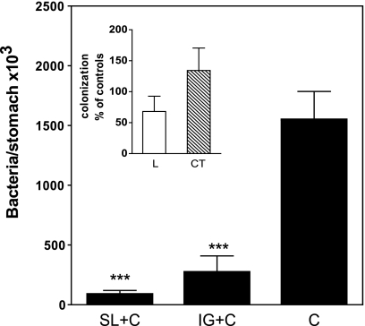
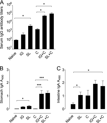
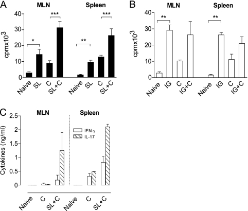
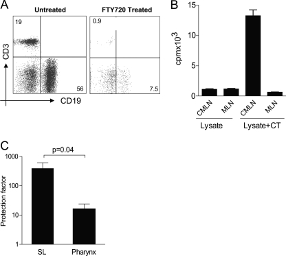
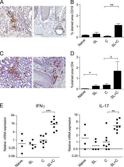
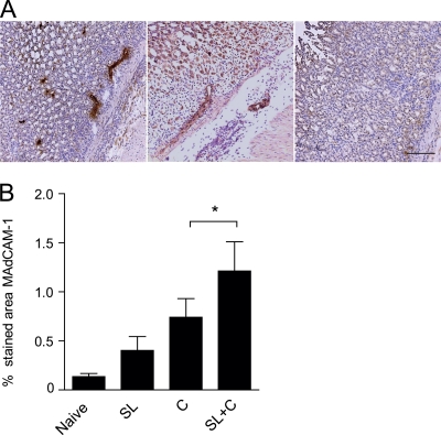
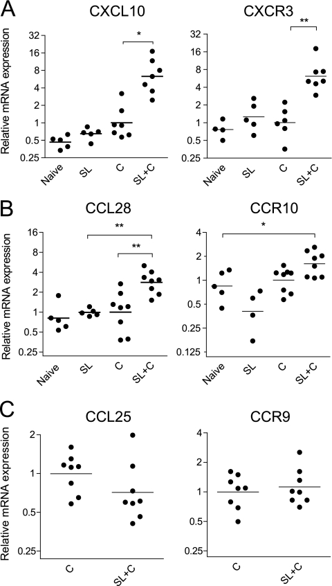
Sublingual (SL) immunization has been described as an effective novel way to induce mucosal immune responses in the respiratory and genital tracts. We examined the potential of SL immunization against Helicobacter pylori to stimulate immune responses in the gastrointestinal mucosa and protect against H. pylori infection. Mice received two SL immunizations with H. pylori lysate antigens and cholera toxin as an adjuvant, and after challenge with live H. pylori bacteria, their immune responses and protection were evaluated, as were immune responses prior to challenge. SL immunization induced enhanced proliferative responses to H. pylori antigens in cervicomandibular lymph nodes and provided at least the same level of immune responses and protection as corresponding intragastric immunization. Protection in SL-immunized mice was associated with strong H. pylori-specific serum IgG and IgA antibody responses in the stomach and intestine, with strong proliferation and gamma interferon (IFN-γ) and interleukin-17 (IL-17) production by spleen and mesenteric lymph node T cells stimulated with H. pylori antigens in vitro, and with increased IFN-γ and IL-17 gene expression in the stomach compared to levels in infected unimmunized mice. Immunohistochemical studies showed enhanced infiltration of CD4(+) T cells and CD19(+) B cells into the H. pylori-infected stomach mucosa of SL-immunized but not unimmunized H. pylori-infected mice, which coincided with increased expression of the mucosal addressin cell adhesion molecule (MAdCAM-1) and T and B cell-attracting chemokines CXCL10 and CCL28. We conclude that, in mice, SL immunization can effectively induce protection against H. pylori infection in association with strong T and B cell infiltration into the stomach.


Figures

FIG. 1.
FIG. 1.
Sublingual immunization protects against H. pylori infection in mice. The data are mean H. pylori CFU + standard error of the mean (SEM) at 4 weeks after challenge in the stomachs of mice that were immunized twice with H. pylori lysate antigens and CT via the sublingual (SL) or intragastric (IG) route, followed 1 week later by challenge with live H. pylori (SL+C and IG+C, respectively) in comparison with concurrently challenged unimmunized mice serving as infection controls (C). Data are from two or three independent experiments with 5 to 10 mice per group in each experiment and show significant differences between both SL- and IG-immunized groups compared to infected controls. ***, P < 0.001. The inset shows colonization of H. pylori in mice that were similarly SL immunized with either lysate antigens alone (L) or CT alone (CT) compared to unimmunized concurrently challenged mice. Data are expressed as mean (+SEM) percentage of bacteria per stomach in individual immunized mice in relation to the mean number of bacteria in infection controls.
FIG. 2.
FIG. 2.
Sublingual immunization induces H. pylori-specific serum antibody responses as well as a local H. pylori-specific IgA antibody response in the stomach mucosa both before and after challenge with live bacteria. Mice tested were as follows: age-matched unimmunized, uninfected C57BL/6 mice served as naïve controls; SL and IG are mice immunized by the SL or IG route and tested before challenge with H. pylori bacteria to study the effect of immunization alone on systemic and local antibody responses; C are unimmunized mice that were challenged with H. pylori and tested 4 weeks later; and SL+C and IG+C are mice that were immunized twice with H. pylori lysate antigens and CT via the sublingual (SL) or intragastric (IG) route, followed 1 week later by challenge with live H. pylori and tested 4 weeks after challenge. (A) Serum antibodies. H. pylori-specific serum IgG antibody titers in the groups of mice indicated were determined by ELISA. Data are representative of one of two independent experiments giving similar results. The bars represent mean + SEM antibody titers, *, P < 0.05. (B and C) IgA antibodies in stomachs and intestinal mucosae. Saponin extracts of stomach (B) and small intestinal (C) mucosa were analyzed for H. pylori-specific IgA antibodies by ELISA. Data represent mean (+SEM) absorbance values at 490 nm (A490) from one of two independent experiments giving similar results. *, P < 0.05; ***, P < 0.001.
FIG. 3.
FIG. 3.
Induction of cellular immune responses to H. pylori in sublingual (SL)-immunized and infected mice. (A and B) In vitro proliferative responses to H. pylori antigens by mesenteric lymph nodes (MLN) and spleen cells after SL (A) compared to IG (B) immunization. Age-matched unimmunized, uninfected C57BL/6 mice served as naïve controls. SL or IG indicate mice that were immunized but not challenged to study the effect of immunization on spleen and MLN proliferative responses. Group C are mice that were left unimmunized, challenged with live H. pylori, and tested 4 weeks later, serving as infection controls, while SL+C and IG+C are mice that were immunized twice SL or IG with H. pylori lysate antigens and CT, followed 1 week later by challenge, and tested 4 weeks after challenge. Data show the incorporation of radioactive thymidine added during the last 6 to 8 h of a 96-h culture, expressed as mean counts per minute (cpm) + SEM, *, P < 0.05; **, P < 0.01; ***, P < 0.001. (C) IFN-γ and IL-17 cytokine levels in cell culture supernatants of cells similar to those for panel A collected 96 h after stimulation, expressed as mean cytokine concentrations (ng per ml) + SEM. Data are from two independent experiments with a pool of five or six mice per group in each experiment.
FIG. 4.
FIG. 4.
CMLN are the primary inductive site after SL immunization. (A) FTY720 treatment prevents egress of T and B cells as evidenced by a reduced frequency of CD3 and CD19 cells in circulation as analyzed by flow cytometry. Numbers represent frequencies of CD3+ T cells and CD19+ B cells in the live lymphocyte gate. (B) Mice were immunized SL with lysate antigens alone or together with CT and treated with FTY720. Single-cell suspensions were prepared from the CMLN and MLN at 5 days after SL immunization and restimulated in vitro with H. pylori antigens. Bars represent mean cpm + SEM of incorporated radioactive thymidine. Data are representative of one of two independent experiments giving similar results. (C) Mice were immunized either SL or in the posterior pharynx and challenged intragastrically with H. pylori bacteria. The CFU recovered from the mouse stomach was used to calculate the protection factors as described in Materials and Methods.
FIG. 5.
FIG. 5.
Increased expression of CD19+ B cells, CD4+ T cells, IFN-γ, and IL-17 in the stomach mucosa of SL-immunized mice. Age-matched unimmunized, uninfected C57BL/6 mice served as naïve controls. SL are mice that were immunized but not challenged to study the effect of immunization alone on the infiltration of T and B cells and cytokine gene expression in the stomach. Group C are mice that were left unimmunized and concurrently challenged and tested 4 weeks later, serving as infection controls, while SL+C are mice that were immunized twice SL with H. pylori lysate antigens and CT followed 1 week later by challenge with live H. pylori and tested 4 weeks after challenge. (A and C) Representative staining patterns for CD19+ B cells and CD4+ T cells in SL-immunized and challenged mice (left) or unimmunized challenged mice (right). Magnification, ×400; bar, 50 μm. (B and D) Cumulative results showing the percentage (mean + SEM) of stained stomach mucosa. Combined data from two experiments are shown, with three or four animals per group and experiment. **, P < 0.01; *, P < 0.05. (E) Expression of cytokine genes. Stomach tissue samples from the same mice as for panels B and D were immediately preserved in RNAlater. Isolated RNA samples were converted into cDNA and analyzed by RT-PCR for IFN-γ and IL-17 transcript levels. The dotted line represents the detection limit. Combined data from two experiments are shown, with n = 2 to 4 animals per group and experiment. ***, P < 0.001; **, P < 0.01.
FIG. 6.
FIG. 6.
Increased MAdCAM-1 expression in the stomachs of SL-immunized and H. pylori-infected mice. (A) MAdCAM-1 staining with characteristic endothelial localization in the stomach tissue of SL-immunized and infected mice (left) and infection control (middle) or isotype control staining (right). Magnification, ×200; bar, 100 μm. (B) Results showing the mean percentage of stomach mucosa specifically stained in the different treatment groups + SEM. Age-matched unimmunized, uninfected mice served as naïve controls. SL are mice that were immunized but not challenged to study the effect of immunization alone on MAdCAM-1 expression in the stomach mucosa. C are mice that were left unimmunized and concurrently challenged, serving as infection controls, while SL+C are mice that were immunized twice SL with H. pylori lysate antigens and CT followed 1 week later by challenge with live H. pylori. Combined data from two experiments are shown, with n = 2 or 3 animals per group and experiment, *, P < 0.05.
FIG. 7.
FIG. 7.
RT-PCR analysis shows increased expression of the CXCL10 (IP-10), CCL28 (MEC), CXCR3, and CCR10 genes but not the CCL25 and CCR9 genes in stomach mucosa of SL-immunized mice and H. pylori infected mice. (A) CXCL10 (left) and CXCR3 (right). (B) CCL28 (left) and CCR10 (right). (C) CCL25 (left) and CCR9 (right). Gene expression in the stomach mucosa of mice from different treatment groups was calculated as described in Materials and Methods. Age-matched unimmunized, uninfected C57BL/6 mice served as naïve controls. SL+C are mice that were immunized twice SL with H. pylori lysate antigens and CT, followed 1 week later by challenge with live H. pylori. Group C are mice that were left unimmunized and concurrently challenged, serving as infection controls, while SL and IG are mice that were immunized but not challenged to study the effect of immunization alone on chemokine and chemokine receptor gene expression in the stomach mucosa Combined data from two experiments are shown; n = 2 or 3 animals per group. *, P < 0.05; **, P < 0.01.

Similar articles

Cited by

References

    1. Akhiani, A. A., J. Pappo, Z. Kabok, K. Schon, W. Gao, L. E. Franzen, and N. Lycke. 2002. Protection against Helicobacter pylori infection following immunization is IL-12-dependent and mediated by Th1 cells. J. Immunol. 169:6977-6984. - PubMed
    1. Akhiani, A. A., K. Schon, L. E. Franzen, J. Pappo, and N. Lycke. 2004. Helicobacter pylori-specific antibodies impair the development of gastritis, facilitate bacterial colonization, and counteract resistance against infection. J. Immunol. 172:5024-5033. - PubMed
    1. Bhuiyan, T. R., A. Saha, A. Lundgren, F. Qadri, and A. M. Svennerholm. Immune responses to Helicobacter pylori infection in Bangladeshi children during their first two years of life and relation between maternal antibodies and onset of infection. J. Infect. Dis., in press. - PubMed
    1. Blaser, M. J., and J. C. Atherton. 2004. Helicobacter pylori persistence: biology and disease. J. Clin. Invest. 113:321-333. - PMC - PubMed
    1. Butcher, E. C., M. Williams, K. Youngman, L. Rott, and M. Briskin. 1999. Lymphocyte trafficking and regional immunity. Adv. Immunol. 72:209-253. - PubMed

Publication types

MeSH terms