. 2010 Oct;78(10):4268-75.
doi: 10.1128/IAI.00469-10. Epub 2010 Aug 9.

Platelet activation and biofilm formation by Aerococcus urinae, an endocarditis-causing pathogen

Affiliations

Platelet activation and biofilm formation by Aerococcus urinae, an endocarditis-causing pathogen

Oonagh Shannon et al. Infect Immun. 2010 Oct.

Abstract

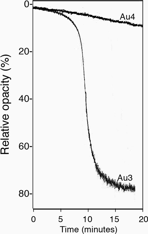
The Gram-positive bacterium Aerococcus urinae can cause infectious endocarditis (IE) in older persons. Biofilm formation and platelet aggregation are believed to contribute to bacterial virulence in IE. Five A. urinae isolates from human blood were shown to form biofilms in vitro, and biofilm formation was enhanced by the presence of human plasma. Four of the A. urinae isolates caused platelet aggregation in platelet-rich plasma from healthy donors. The Au3 isolate, which induced platelet aggregation in all donors, also activated platelets, as determined by flow cytometry. Platelet aggregation was dependent on bacterial protein structures and on platelet activation since it was sensitive to both trypsin and prostaglandin E(1). Plasma proteins at the bacterial surface were needed for platelet aggregation; and roles of the complement system, fibrinogen, and immunoglobulin G were demonstrated. Complement-depleted serum was unable to support platelet aggregation by Au3 and complement blockade using compstatin-inhibited platelet activation. Platelet activation by Au3 was inhibited by blocking of the platelet fibrinogen receptor, and this isolate was also shown to bind to radiolabeled fibrinogen. Removal of IgG from platelet-rich plasma by a specific protease inhibited the platelet aggregation induced by A. urinae, and blockade of the platelet FcRγIIa hindered platelet activation induced by Au3. Convalescent-phase serum from a patient with A. urinae IE transferred the ability of the bacterium to aggregate platelets in an otherwise nonresponsive donor. Our results show that A. urinae exhibits virulence strategies of importance for IE.


Figures

FIG. 1.
FIG. 1.
A. urinae biofilm formation is stimulated by plasma. Biofilm formation by A. urinae isolates after 72 h incubation on a plastic surface was determined as the absorbance at 550 nm in the presence of medium (black bars) or medium containing 10% human plasma (gray bars). Biofilm formation was also determined in wells pretreated with human plasma (white bars). The error bars represent the standard deviations from three independent experiments, carried out in triplicate.
FIG. 2.
FIG. 2.
Morphology of the A. urinae biofilm. Scanning electron microscopy was used to visualize biofilms formed by Au1 (A and B) and Au3 (C and D) on glass coverslips in medium (A and C) or in medium containing 10% human plasma (B and D). Bar, 10 μm.
FIG. 3.
FIG. 3.
A. urinae stimulates platelet aggregation. Platelet aggregation in PRP from donor 1 in response to A. urinae isolates Au3 and Au4 was determined using a Chrono-Log aggregometer. The relative light absorption of the sample is shown on the y axis, where 100 represents the absorption of PRP after addition of bacteria and 0 the absorption of PPP. When platelets aggregate the opacity, the light absorbance of the sample decreases.
FIG. 4.
FIG. 4.
Platelet aggregation and activation by Au3 is dependent on the complement system. (A) Au3 was either pretreated with PPP, followed by addition of PRP from donor 1 (curve b), or added to a mixture of PPP and PRP from the same donor (curve a), and aggregation was determined by aggregometry. (B) Washed platelets from donor 1 were added to citrated plasma from the same donor, followed by addition of Au3 bacteria (curve a). Washed platelets were added to serum reconstituted with fibrinogen (curve b) or heat-treated serum reconstituted with fibrinogen (curve c), followed by addition of Au3 bacteria. AP1 was able to induce aggregation of washed platelets in heat-treated serum reconstituted with fibrinogen (curve d).
FIG. 5.
FIG. 5.
Role of platelet fibrinogen receptor and fibrinogen binding by A. urinae. Au3 was added to PRP from the five donors, and after 25 min, platelet activation was determined using flow cytometry. The percentage of the platelet population positive for CD62P (A) and PAC-1 (B) was determined in the presence of buffer alone (white bars) or an antibody to block platelet fibrinogen binding (ReoPro; black bars). (C) The binding of radiolabeled fibrinogen to the A. urinae isolates and to the AP1 strain of S. pyogenes is expressed as the percentage of added fibrinogen associated with the bacterial pellet (n = 3; bars represent standard deviations). (D) The binding of radiolabeled fibrinogen to Au3 (triangles) and to the AP1 strain of S. pyogenes (squares) was determined in the presence of unlabeled fibrinogen. Binding is expressed as a percentage of the binding of radiolabeled fibrinogen alone. Results are from three independent experiments, and the bars represent standard deviations.
FIG. 6.
FIG. 6.
Role of plasma IgG in platelet activation and aggregation by A. urinae. Au3 was added to PRP from the five donors, and after 25 min, platelet activation was determined using flow cytometry. The percentage of the platelet population positive for CD62P (A) and PAC-1 (B) was determined in the presence of buffer alone (white bars) or an antibody to block platelet Ig binding (AT10; black bars). (C) PRP from donor 1 was pretreated with the IgG-degrading enzyme IdeS or buffer, followed by addition of collagen or Au3 bacteria. Collagen induced rapid aggregation in PRP pretreated with IdeS (curve a) or buffer (curve b). Au3 bacteria failed to induce aggregation in PRP pretreated with IdeS (curve c), whereas aggregation occurred in PRP treated with buffer alone (curve d). (D) Aggregation of platelets in PRP from donor 1 in response to Au1 and Au3 was determined. In the presence of 25 μl of serum from donor 1, platelets did not aggregate in response to Au1 (curve a), whereas aggregation occurred in response to Au3 (curve d). In the presence of 25 μl of serum from the patient infected with Au1, the platelets aggregated in response to Au1 (curve b). Patient serum preincubated with IdeS could not mediate platelet aggregation in response to Au1 (curve c).

References

    1. Aguirre, M., and M. D. Collins. 1992. Phylogenetic analysis of some Aerococcus-like organisms from urinary tract infections: description of Aerococcus urinae sp. nov. J. Gen. Microbiol. 138:401-405. - PubMed
    1. Åkesson, P., K. H. Schmidt, J. Cooney, and L. Björck. 1994. M1 protein and protein H: IgGFc- and albumin-binding streptococcal surface proteins encoded by adjacent genes. Biochem. J. 300(Pt 3):877-886. - PMC - PubMed
    1. Astudillo, L., L. Sailler, L. Porte, J. C. Lefevre, P. Massip, and E. Arlet-Suau. 2003. Spondylodiscitis due to Aerococcus urinae: a first report. Scand. J. Infect. Dis. 35:890-891. - PubMed
    1. Bayer, A. S., D. Cheng, M. R. Yeaman, G. R. Corey, R. S. McClelland, L. J. Harrel, and V. G. Fowler, Jr. 1998. In vitro resistance to thrombin-induced platelet microbicidal protein among clinical bacteremic isolates of Staphylococcus aureus correlates with an endovascular infectious source. Antimicrob. Agents Chemother. 42:3169-3172. - PMC - PubMed
    1. Bensing, B. A., J. A. Lopez, and P. M. Sullam. 2004. The Streptococcus gordonii surface proteins GspB and Hsa mediate binding to sialylated carbohydrate epitopes on the platelet membrane glycoprotein Ibα. Infect. Immun. 72:6528-6537. - PMC - PubMed

Publication types

LinkOut - more resources