

Invasion and multiplication of Helicobacter pylori in gastric epithelial cells and implications for antibiotic resistance
- PMID: 20696835
- PMCID: PMC2950335
- DOI: 10.1128/IAI.00524-10
Invasion and multiplication of Helicobacter pylori in gastric epithelial cells and implications for antibiotic resistance
Abstract
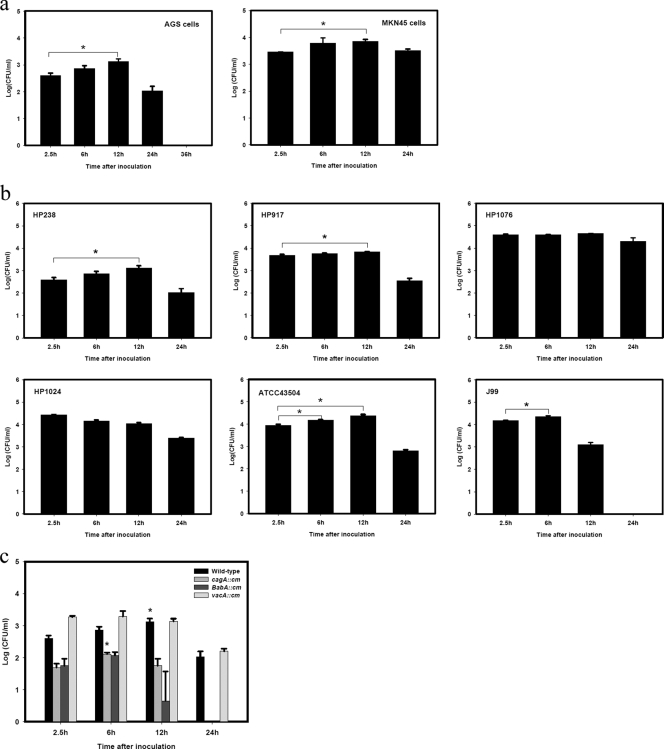
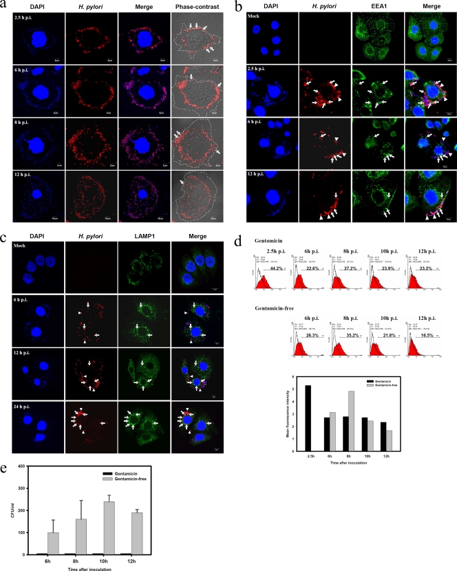
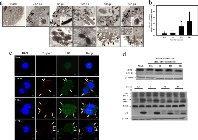
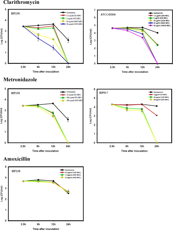
Helicobacter pylori is a Gram-negative, spiral-shaped bacterium that infects more than 50% of the human population and can cause gastritis, peptic ulcer, or gastric malignancies. It is generally viewed as an extracellular microorganism. In a gentamicin protection assay on AGS or MKN45 cells, H. pylori could invade the epithelial cells and multiply within double-layer vesicles either on the plasma membrane or in the cytoplasm. A 5-fold increase in the number of bacteria was recultured from the infected cells at 12 h, compared with the number of invading cells at 2.5 h postinfection. The autophagic vesicles induced by H. pylori are the sites of replication and also of the degradation of the replicating bacteria after fusion with lysosomes. Many H. pylori bacteria in coccoid form associated with the plasma membrane can be released into culture. Only cell-penetrating antibiotics can enhance the intracellular killing of the replicating bacteria. The multiplication of H. pylori within cells provides a niche for its resistance to antibacterial therapy and has a significant impact on its biological life cycle.
Figures
Similar articles
-
Helicobacter pylori: an invading microorganism? A review.FEMS Immunol Med Microbiol. 2003 May 25;36(3):117-26. doi: 10.1016/S0928-8244(03)00020-8. FEMS Immunol Med Microbiol. 2003. PMID: 12738380 Review.
-
The internalization of Helicobacter pylori plays a role in the failure of H. pylori eradication.Helicobacter. 2017 Feb;22(1). doi: 10.1111/hel.12324. Epub 2016 Jun 10. Helicobacter. 2017. PMID: 27282442
-
Helicobacter pylori: molecular basis for colonization and survival in gastric environment and resistance to antibiotics. A short review.Infect Dis (Lond). 2019 Jun;51(6):399-408. doi: 10.1080/23744235.2019.1588472. Epub 2019 Mar 25. Infect Dis (Lond). 2019. PMID: 30907202 Review.
-
Helicobacter pylori enter and survive within multivesicular vacuoles of epithelial cells.Cell Microbiol. 2002 Oct;4(10):677-90. doi: 10.1046/j.1462-5822.2002.00222.x. Cell Microbiol. 2002. PMID: 12366404
-
Helicobacter pylori and antimicrobial resistance: molecular mechanisms and clinical implications.Lancet Infect Dis. 2006 Nov;6(11):699-709. doi: 10.1016/S1473-3099(06)70627-2. Lancet Infect Dis. 2006. PMID: 17067919 Review.
Cited by
-
The impact of autophagic processes on the intracellular fate of Helicobacter pylori: more tricks from an enigmatic pathogen?Autophagy. 2013 May;9(5):639-52. doi: 10.4161/auto.23782. Epub 2013 Feb 8. Autophagy. 2013. PMID: 23396129 Free PMC article. Review.
-
Molecular mechanism of Helicobacter pylori-induced autophagy in gastric cancer.Oncol Lett. 2019 Dec;18(6):6221-6227. doi: 10.3892/ol.2019.10976. Epub 2019 Oct 10. Oncol Lett. 2019. PMID: 31788098 Free PMC article. Review.
-
Towards Fluorescence In Vivo Hybridization (FIVH) Detection of H. pylori in Gastric Mucosa Using Advanced LNA Probes.PLoS One. 2015 Apr 27;10(4):e0125494. doi: 10.1371/journal.pone.0125494. eCollection 2015. PLoS One. 2015. PMID: 25915865 Free PMC article.
-
Nonhelical Helicobacter pylori Mutants Show Altered Gland Colonization and Elicit Less Gastric Pathology than Helical Bacteria during Chronic Infection.Infect Immun. 2019 Jun 20;87(7):e00904-18. doi: 10.1128/IAI.00904-18. Print 2019 Jul. Infect Immun. 2019. PMID: 31061142 Free PMC article.
-
Helicobacter pylori inhibits autophagic flux and promotes its intracellular survival and colonization by down-regulating SIRT1.J Cell Mol Med. 2021 Apr;25(7):3348-3360. doi: 10.1111/jcmm.16411. Epub 2021 Feb 28. J Cell Mol Med. 2021. PMID: 33641223 Free PMC article.
References
-
- Allen, L. A. 2007. Phagocytosis and persistence of Helicobacter pylori. Cell Microbiol. 9:817-828. - PubMed
-
- Amieva, M. R., N. R. Salama, L. S. Tompkins, and S. Falkow. 2002. Helicobacter pylori enter and survive within multivesicular vacuoles of epithelial cells. Cell Microbiol. 4:677-690. - PubMed
-
- Deretic, V. 2006. Autophagy as an immune defense mechanism. Curr. Opin. Immunol. 18:1-8. - PubMed
Publication types
MeSH terms
Substances
LinkOut - more resources
Full Text Sources
Medical
