. 2010 Oct;30(20):4940-51.
doi: 10.1128/MCB.00642-10. Epub 2010 Aug 9.

Dosage-sensitive regulation of cohesin chromosome binding and dynamics by Nipped-B, Pds5, and Wapl

Affiliations

Dosage-sensitive regulation of cohesin chromosome binding and dynamics by Nipped-B, Pds5, and Wapl

Maria Gause et al. Mol Cell Biol. 2010 Oct.

Abstract

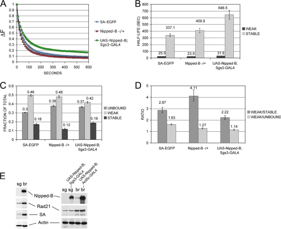
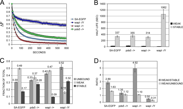
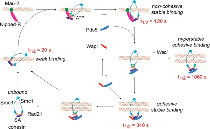
The cohesin protein complex holds sister chromatids together to ensure proper chromosome segregation upon cell division and also regulates gene transcription. Partial loss of the Nipped-B protein that loads cohesin onto chromosomes, or the Pds5 protein required for sister chromatid cohesion, alters gene expression and organism development, without affecting chromosome segregation. Knowing if a reduced Nipped-B or Pds5 dosage changes how much cohesin binds chromosomes, or the stability with which it binds, is critical information for understanding how cohesin regulates transcription. We addressed this question by in vivo fluorescence recovery after photobleaching (FRAP) with Drosophila salivary glands. Cohesin, Nipped-B, and Pds5 all bind chromosomes in both weak and stable modes, with residence half-lives of some 20 seconds and 6 min, respectively. Reducing the Nipped-B dosage decreases the amount of stable cohesin without affecting its chromosomal residence time, and reducing the Pds5 dosage increases the amount of stable cohesin. This argues that Nipped-B and Pds5 regulate transcription by controlling how much cohesin binds DNA in the stable mode, and not binding affinity. We also found that Nipped-B, Pds5, and the Wapl protein that interacts with Pds5 all play unique roles in cohesin chromosome binding.


Figures

FIG. 1.
FIG. 1.
Expression of EGFP fusion cohesion proteins. (A) SA-EGFP and EGFP-Smc1 interact with cohesin. Nuclear extracts of control cells (+) or ML-DmD8 cells expressing SA-EGFP or Kc cells expressing EGFP-Smc1 were immunoprecipitated (IP) with anti-Smc1 or anti-Rad21 and subjected to Western blot analysis with anti-SA or anti-Smc1, showing that the fusion proteins interact with cohesin. (B) Whole-cell extracts of 3rd-instar control (+) brains or brains expressing SA-EGFP or Nipped-B-EGFP were subjected to Western blot analysis with the indicated antibodies.
FIG. 2.
FIG. 2.
EGFP-Smc1 dynamics in Kc cells. (A) FACS analysis of Kc cells before or after copper (Cu2+) induction of EGFP-Smc1 expression. (B) FRAP recovery of EGFP-Smc1 after photobleaching in group I (G1) and group II (G2) cells, which represent some 30% and 70% of cells, respectively. The difference in fluorescence (ΔF) between the unbleached half and the bleached half of the nucleus is plotted versus the time postbleaching. We assume that group I cells are in G1 and group II are in G2. (C) Half-lives of weak- and stable-binding modes of EGFP-Smc1 chromosome, determined with a two-binding mode model. (D) Distribution of EGFP-Smc1 into unbound, weak binding, and stable binding. Unbound EGFP-Smc1 was determined by measuring the loss of fluorescence in the unbleached half of the nucleus. (E) Ratios of weak-binding EGFP-Smc1 to stable-binding and unbound EGFP-Smc1. Error bars are standard errors of the mean.
FIG. 3.
FIG. 3.
SA-EGFP, EGFP-Smc1, Nipped-B-EGFP, and Pds5-EGFP dynamics in 3rd-instar salivary gland cells. (A) FRAP recovery curves for SA-EGFP, EGFP-Smc1, Nipped-B-EGFP, and Pds5-EGFP. The SA-EGFP, EGFP-Smc1, and Nipped-B-EGFP curves are overlapping, while Pds5-EGFP shows faster recovery. (B) Chromosomal half-lives for weak- and strong-binding modes of SA-EGFP, EGFP-Smc1, Nipped-B-EGFP, and Pds5-EGFP. (C) Distribution of SA-EGFP, EGFP-Smc1, Nipped-B-EGFP, and Pds5-EGFP into unbound and weakly and strongly bound fractions. (D) Ratio of weak-binding SA-EGFP, EGFP-Smc1, Nipped-B-EGFP, and Pds5-EGFP to stable binding and unbound forms. Error bars are standard errors of the mean.
FIG. 4.
FIG. 4.
SA-EGFP dynamics in G- and S-phase salivary gland cells. (A) (Top) A salivary gland in which S phase has been induced by the hs-cyclin E transgene. The left column (green) shows EGFP-SA fluorescence, the middle (red) shows EdU staining, which indicates DNA synthesis, and the right shows DNA DAPI staining. (Bottom) A control salivary gland subjected to the same heat shock without the hs-cyclin E transgene. (B) FRAP recovery curves for G-phase nuclei in control salivary glands (blue diamonds) and S-phase nuclei induced by cyclin E expression (red circles). (C) Half-lives of weak and stable SA-EGFP chromosome binding in G- and S-phase nuclei. (D) Distribution of SA-EGFP into unbound, weakly bound, and strongly bound forms in G- and S-phase nuclei. (E) Ratios of weakly bound SA-EGFP to the stably bound and unbound.
FIG. 5.
FIG. 5.
Effects of Nipped-B dosage on SA-EGFP dynamics in salivary gland cells. (A) FRAP recovery curves for SA-EGFP in wild-type salivary cells (SA-EGFP) and Nipped-B407/+ mutants (Nipped-B −/+) and with overexpressed Nipped-B (UAS-Nipped-B; Sgs3-Gal4). (B) Chromosomal half-lives for weak- and strong-binding SA-EGFP. (C) Distribution of SA-GFP into unbound, weakly bound, and strongly bound forms. (D) Ratio of weak-binding SA-EGFP to stably bound and unbound. (E) Western blots of salivary glands (sg) and brains (br) comparing expression of Nipped-B, Rad21, and SA, relative to actin, in sg and br. All lanes on left are the same Western blot. The right lanes compare levels of expression in sg without and with the UAS-Nipped-B; Sgs3-GAL4 overexpression transgenes.
FIG. 6.
FIG. 6.
Effects of Pds5 and Wapl dosages on SA-EGFP dynamics in salivary gland cells. (A) SA-EGFP FRAP recovery curves for wild-type (SA-EGFP), pds5e3/+ (pds5 −/+), wapl2/+ (wapl −/+), and wapl2/Y (wapl −/Y) salivary cells. (B) Chromosomal half-lives for weak and stable SA-EGFP. (C) Distribution of SA-EGFP into unbound, weakly bound, and strongly bound fractions. (D) Ratio of weakly bound SA-EGFP to stably bound and unbound. Error bars are standard errors of the mean.
FIG. 7.
FIG. 7.
Models for the roles of Nipped-B, Pds5, and Wapl in regulating cohesin chromosome binding and dynamics. We hypothesize that weak-binding cohesin with a chromosomal half-life (t1/2) of approximately 20 s associates with DNA without encircling it. The Nipped-B/Mau-2 complex binds DNA and facilitates opening of the cohesin ring to allow it to encircle DNA. With excess Pds5, and sister chromatids, this is rapidly converted to stable “cohesive” cohesin with a measured half-life of approximately 340 s. The cohesive mode could consist of a single cohesin ring encircling two chromatids as illustrated or two interacting cohesin rings that each encircle a chromatid. The Pds5-Wapl complex can remove stable cohesin to generate unbound cohesin or weakly bound cohesin. We postulate that free Pds5 interferes with cohesin loading by Nipped-B/Mau-2, perhaps by competing for binding to cohesin. The Pds5-Wapl complex does not inhibit loading, but a decrease in Wapl increases the amount of free Pds5, thereby reducing loading. In the absence of Wapl, free Pds5 still makes cohesive cohesin, which binds with a half-life of approximately 1,060 s.

Similar articles

Cited by

References

    1. Arumugam, P., S. Gruber, K. Tanaka, C. H. Haering, K. Mechtler, and K. Nasmyth. 2003. ATP hydrolysis is required for cohesin's association with chromosomes. Curr. Biol. 13:1941-1953. - PubMed
    1. Bernard, P., J. Drogat, J. F. Maure, S. Dheur, S. Vaur, S. Genier, and J. P. Javerzat. 2006. A screen for cohesion mutants uncovers Ssl3, the fission yeast counterpart of the cohesin loading factor Scc4. Curr. Biol. 16:875-881. - PubMed
    1. Bunch, T. A., Y. Grinblat, and L. S. Goldstein. 1988. Characterization and use of the Drosophila metallothionein promoter in cultured Drosophila melanogaster cells. Nucleic Acids Res. 16:1043-1061. - PMC - PubMed
    1. Ciosk, R., M. Shirayama, A. Shevchenko, T. Tanaka, A. Toth, A. Shevchenko, and K. Nasmyth. 2000. Cohesin's binding to chromosomes depends on a separate complex consisting of Scc2 and Scc4 proteins. Mol. Cell 5:243-254. - PubMed
    1. Deardorff, M. A., M. Kaur, D. Yaeger, A. Rampuria, S. Korolev, J. Pie, C. Gil-Rodríguez, M. Arnedo, B. Loeys, A. D. Kline, M. Wilson, K. Lillquist, V. Siu, F. J. Ramos, A. Musio, L. S. Jackson, D. Dorsett, and I. D. Krantz. 2007. Mutations in cohesin complex members SMC3 and SMC1A cause a mild variant of Cornelia de Lange syndrome with predominant mental retardation. Am. J. Hum. Genet. 80:485-494. - PMC - PubMed

Publication types

MeSH terms

LinkOut - more resources