. 2010 Aug 24;107(34):15157-62.
doi: 10.1073/pnas.1003822107. Epub 2010 Aug 9.

TCF4 and CDX2, major transcription factors for intestinal function, converge on the same cis-regulatory regions

Affiliations

TCF4 and CDX2, major transcription factors for intestinal function, converge on the same cis-regulatory regions

Michael P Verzi et al. Proc Natl Acad Sci U S A. .

Abstract

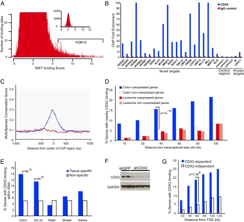
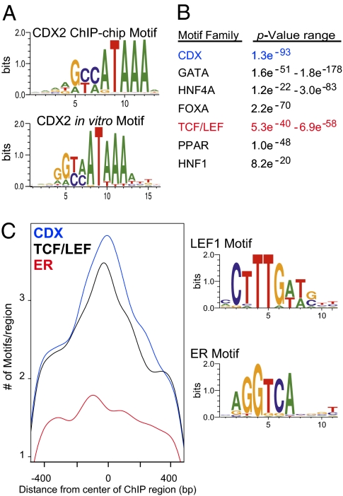
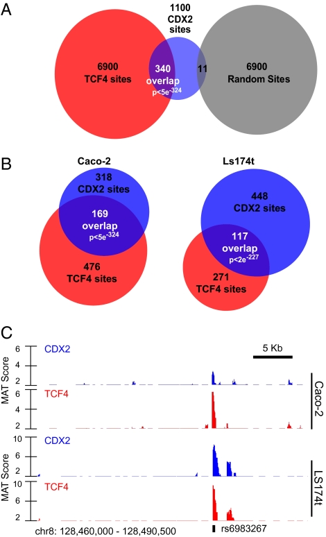
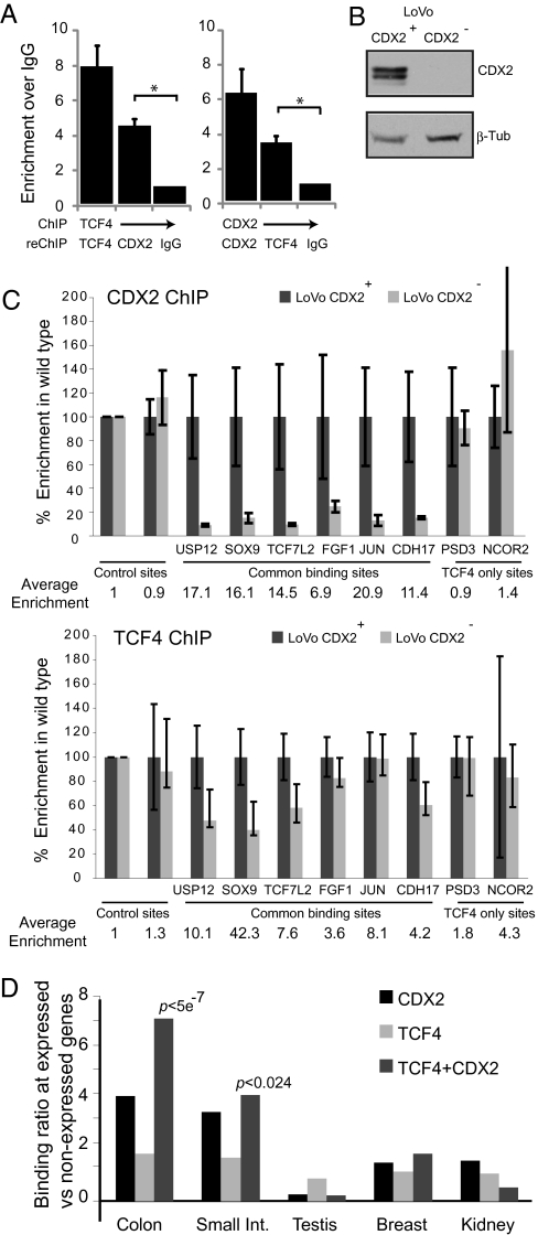
Surprisingly few pathways signal between cells, raising questions about mechanisms for tissue-specific responses. In particular, Wnt ligands signal in many mammalian tissues, including the intestinal epithelium, where constitutive signaling causes cancer. Genome-wide analysis of DNA cis-regulatory regions bound by the intestine-restricted transcription factor CDX2 in colonic cells uncovered highly significant overrepresentation of sequences that bind TCF4, a transcriptional effector of intestinal Wnt signaling. Chromatin immunoprecipitation confirmed TCF4 occupancy at most such sites and co-occupancy of CDX2 and TCF4 across short distances. A region spanning the single nucleotide polymorphism rs6983267, which lies within a MYC enhancer and confers colorectal cancer risk in humans, represented one of many co-occupied sites. Co-occupancy correlated with intestine-specific gene expression and CDX2 loss reduced TCF4 binding. These results implicate CDX2 in directing TCF4 binding in intestinal cells. Co-occupancy of regulatory regions by signal-effector and tissue-restricted transcription factors may represent a general mechanism for ubiquitous signaling pathways to achieve tissue-specific outcomes.


Conflict of interest statement

The authors declare no conflict of interest.

Figures

Fig. 1.
Fig. 1.
CDX2 binding in the Caco-2 genome. (A) Histogram showing CDX2 ChIP-chip binding sites identified by MAT (15) at different MAT scores. Sites outside the normal distribution were assigned confidence scores to impute FDRs. (B) Representative validation of unique sites in independent CDX2 IPs, followed by quantitative PCR (qPCR) analysis of selected genomic regions. Mean fold-enrichment in ChIP is expressed relative to input or IgG IPs and control regions. Sites are named according to the nearest locus; genome coordinates are listed in Table S1. (C) Composite plot of multispecies conservation at CDX2-occupied regions, showing peak conservation at the centers (blue); random regions (red) show background conservation. (D) Oncomine was used to identify genes coexpressed with CDX2 in cancer samples (16). Genes coexpressed with CDX2 in colon tumors are more likely to have nearby CDX2 binding than genes not coexpressed with CDX2; gene coexpression in leukemia showed no correlation with intestinal CDX2 binding. (E) RNA expression data from 55 mouse tissues (18) were correlated with CDX2 occupancy within 60-kb windows. Human orthologs of genes highly expressed in mouse small and large intestine are much more likely to have nearby CDX2 occupancy in Caco-2 cells than genes not expressed in mouse intestine. (F) Immunoblot demonstration of CDX2 depletion in Caco-2 cells following lentiviral delivery of shCDX2. (G) Genes dysregulated upon CDX2 depletion are more likely to have nearby CDX2 binding sites than unaffected genes.
Fig. 2.
Fig. 2.
Computational prediction that intestinal transcription factors co-occupy CDX2 binding regions. (A) Position-weighted matrices demonstrating that the most centered sequence motif identified de novo (P < 1e−30, Upper) within CDX2-occupied regions closely resembles the consensus motif attributed to CDX2 (Lower) (19). (B) TRANSFAC motifs identified using CEAS (21), with the corresponding P value for enrichment in CDX2-bound regions over the genomic background. Each of these motifs is associated with transcription factors known to regulate intestinal genes. (C) Density plots of motif occurrences at CDX2 ChIP regions shows disproportionate enrichment of CDX2 and TCF/LEF motifs at the centers of CDX2-occupied regions, suggesting functionality; the estrogen-response element (ERE) is not enriched or centered. Motif matrices used in the analysis are shown on the right.
Fig. 3.
Fig. 3.
The cis-regulatory region co-occupancy by CDX2 and the Wnt-effector transcription factor TCF4. (A) Extensive overlap between 6,900 TCF4-binding regions previously identified (FDR = 0) in Ls174t colon cancer cells (27) and CDX2-occupied regions in Caco-2. Nearly 1 in 3 CDX2-binding sites (FDR = 0) falls within a TCF4-occupied region, compared with <1% that overlap with 6,900 random 1-kb regions. (B) TCF4 ChIP-chip on chromosomes 8, 11, and 12 in Caco-2 (Left) and Ls174t (Right) cells revealed TCF4 occupancy of 26% (Ls174T) to 53% (Caco-2) of CDX2-binding regions on these chromosomes. (C) Raw traces of CDX2 (blue) and TCF4 (red) ChIP-chip data in Caco-2 (Upper) and LS174t (Lower) cells, demonstrating precise co-occupancy at an upstream MYC enhancer that encompasses the colon cancer risk SNP rs6983267.
Fig. 4.
Fig. 4.
Demonstration and functional significance of cis-regulatory region co-occupancy by CDX2 and TCF4. (A) Sequential ChIP with antibody against TCF4 followed by antibody against CDX2 and vice versa reveals the presence of both transcription factors on the same chromatin binding region near EPHB2. The chromatin eluted from the first ChIP was immunoprecipitated again (reChIP) with CDX2, TCF4, or mouse or goat IgG controls. Enrichment is calculated based upon qPCR relative to the IgG control, which is set to 1. Enrichments are indicated with SEM and are the results of three independent experiments. The asterisk represents P < 0.001 (t test). Similar results at a region near ADRA2C are shown in Fig. S4D. (B) Immunoblot evidence of CDX2 expression in parental and modified LoVo cells. (C) ChIP analysis of representative individual and co-occupied sites in paired CDX2+ and CDX2 LoVo lines, with fold-enrichment relative to input DNA determined by qPCR. ChIP enrichment is shown relative to the parental, CDX2-expressing LoVo line. The average ChIP enrichment in the parental line for each region is listed below the graph. CDX2 binding showed dependence on CDX2. TCF4 ChIP revealed that binding at certain co-occupied sites also depends on the presence of CDX2, whereas binding at sites without demonstrable CDX2 occupancy does not. Sites are named according to the nearest locus and genome coordinates are listed in Table S1. (D) Comparison of the ratios of tissue-specific versus nonspecific loci bound by each transcription factor. TCF4- and independent CDX2-binding sites show significant but relatively modest correlation with transcripts expressed in the mouse intestine, whereas co-occupancy of CDX2 and TCF4 is much better associated with intestinal gene expression. Breast, kidney, and testis transcripts showed no association with TCF4 or CDX2 binding near expressed genes.

Similar articles

Cited by

References

    1. James R, Kazenwadel J. Homeobox gene expression in the intestinal epithelium of adult mice. J Biol Chem. 1991;266:3246–3251. - PubMed
    1. Suh E, Traber PG. An intestine-specific homeobox gene regulates proliferation and differentiation. Mol Cell Biol. 1996;16:619–625. - PMC - PubMed
    1. Mutoh H, et al. Conversion of gastric mucosa to intestinal metaplasia in Cdx2-expressing transgenic mice. Biochem Biophys Res Commun. 2002;294:470–479. - PubMed
    1. Silberg DG, et al. Cdx2 ectopic expression induces gastric intestinal metaplasia in transgenic mice. Gastroenterology. 2002;122:689–696. - PubMed
    1. Liu T, et al. Regulation of Cdx2 expression by promoter methylation, and effects of Cdx2 transfection on morphology and gene expression of human esophageal epithelial cells. Carcinogenesis. 2007;28:488–496. - PubMed

Publication types

MeSH terms

Associated data