. 2010 Aug 24;107(34):15287-92.
doi: 10.1073/pnas.0911451107. Epub 2010 Aug 9.

Vinca drug components accumulate exclusively in leaf exudates of Madagascar periwinkle

Affiliations

Vinca drug components accumulate exclusively in leaf exudates of Madagascar periwinkle

Jonathan Roepke et al. Proc Natl Acad Sci U S A. .

Abstract

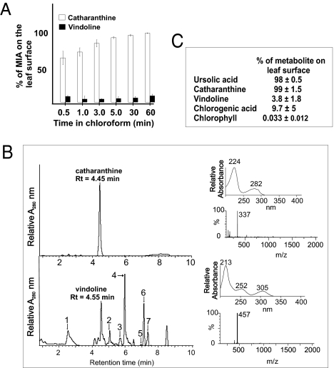
The monoterpenoid indole alkaloids (MIAs) of Madagascar periwinkle (Catharanthus roseus) continue to be the most important source of natural drugs in chemotherapy treatments for a range of human cancers. These anticancer drugs are derived from the coupling of catharanthine and vindoline to yield powerful dimeric MIAs that prevent cell division. However the precise mechanisms for their assembly within plants remain obscure. Here we report that the complex development-, environment-, organ-, and cell-specific controls involved in expression of MIA pathways are coupled to secretory mechanisms that keep catharanthine and vindoline separated from each other in living plants. Although the entire production of catharanthine and vindoline occurs in young developing leaves, catharanthine accumulates in leaf wax exudates of leaves, whereas vindoline is found within leaf cells. The spatial separation of these two MIAs provides a biological explanation for the low levels of dimeric anticancer drugs found in the plant that result in their high cost of commercial production. The ability of catharanthine to inhibit the growth of fungal zoospores at physiological concentrations found on the leaf surface of Catharanthus leaves, as well as its insect toxicity, provide an additional biological role for its secretion. We anticipate that this discovery will trigger a broad search for plants that secrete alkaloids, the biological mechanisms involved in their secretion to the plant surface, and the ecological roles played by them.


Conflict of interest statement

The authors declare no conflict of interest.

Figures

Fig. 1.
Fig. 1.
Catharanthine accumulates almost entirely in leaf wax exudates outside of the leaf epidermis, whereas vindoline is found within leaf cells. (A) Measurement of catharanthine and vindoline extracted from the leaf surface was made by dipping them in chloroform for different times. The experiment for each time point was performed in triplicate. The levels of catharanthine and vindoline per whole leaf representing the 100% value were 240 ± 9 μg and 113 ± 6 μg, respectively, in leaf number 3, as seen in Fig. 2. (B) Chloroform-extracted MIAs and methanol-extracted MIAs remaining in leaves after chloroform treatments were measured by UPLC-MS (Upper). MIAs 1 to 7 were tentatively identified by their absorption and mass spectra as serpentine (1, m/z = 349); deacetoxyvindoline (2, m/z = 399); anhydrovinblastine (3, m/z = 794); unknown (4, m/z = 615); 16-methoxytabersonine (5, m/z = 367); unknown (6, m/z = 594); and unknown (7, m/z = 535 and 594). (C) Triplicate measurements of ursolic acid, catharanthine, vindoline, chlorogenic acid, and chlorophyll extracted from the leaf surface by dipping them in chloroform for 1 h. The levels per leaf pair of ursolic acid, catharanthine, vindoline, chlorogenic acid, and chlorophyll representing 100% were 919 ± 90, 271 ± 63, 120 ± 19, 210 ± 18, and 131 ± 17 μg/g fresh weight, respectively.
Fig. 2.
Fig. 2.
Distribution of catharanthine and vindoline in the leaf wax exudate and within leaf cells in leaves of different ages.
Fig. 3.
Fig. 3.
Model for biosynthesis and secretion of secondary metabolites produced in the epidermis of C. roseus leaves. (A) Enrichment of monoterpenoid indole alkaloid pathway enzyme activities in leaf epidermis enriched protein extracts produced by carborundum abrasion compared with those found in whole leaves. The whole leaf–specific activities used to establish the fold enrichment of enzymes in the leaf epidermis were a follows: G10H (184 ± 14 nmol/μg protein), LAMT (0.69 ± 0.02 pmol/μg protein), 16-OMT (1.89 ± 0.05 pmol/μg protein), NMT (0.47 ± 0.01 pmol/μg protein), and DAT (0.37 ± 0.02 pmol/ μg protein). (B) The leaf epidermis is specialized for biosynthesis of flavonoids, the fatty acid components of waxes, triterpenes, and MIAs. Although the 10-hydroxygeraniol required for biosynthesis of secologanin is made in specialized IPAP cells via the methyl–erythritol pathway (MEP) and G10H, this metabolite or another intermediate is transported to the leaf epidermis, where loganic acid is converted to secologanin via LAMT and secologanin synthase (SLS). Within the leaf epidermis, tryptophan is decarboxylated in the cytoplasm to tryptamine by tryptophan decarboxylase (TDC) and strictosidine is formed by the coupling of tryptamine and secologanin in the leaf epidermal vacuole by strictosidine synthase (STR1). Strictosidine is released into the cytoplasm as a reactive aglycone by cytosolic strictosidine β-glucosidase (SGD), where mostly unknown biochemical transformations lead to the production of the three major—Corynanthe, Iboga, or Aspidosperma—backbones of MIAs. A common intermediate is converted to catharanthine or to tabersonine by uncharacterized biochemical reactions. Virtually all the catharanthine is secreted into the leaf cell surface, together with fatty acid components of waxes and the triterpene, ursolic acid. After tabersonine is converted into 16-methoxytabersonine in the leaf epidermis by tabersonine-16-hydroxylase (T16H) and by 2,3-dihydro-3-hydroxy-N-methyltabersonine (16OMT), this metabolite may be secreted into leaf mesophyl cells, where a further uncharacterized oxidation takes place, followed by a N-methylation (NMT) located in chloroplast thylakoids, hydroxylation (D4H), and O-acetylation (DAT) to yield vindoline. The last two reactions in vindoline biosynthesis appear to occur in specialized leaf idioblast and laticifer cells. It is postulated that wounding or herbivory may bring catharanthine and vindoline together to allow the formation of dimeric anticancer MIAs. The dotted lines represent uncharacterized reactions or unknown mechanisms that remain to be documented.

References

    1. Mangeney P, Andriamialisoa RZ, Langlois N, Langlois Y, Potier P. Preparation of vinblastine, vincristine, and leurosidine, antitumor alkaloids from Catharanthus species (Apocynaceae) J Am Chem Soc. 1979;101:2243–2245.
    1. Kutney JP, et al. A highly efficient and commercially important synthesis of the antitumor catharanthus alkaloids vinblastine and leurosidine from catharanthine and vindoline. Heterocycles. 1988;27:1845–1853.
    1. Kuehne ME, Matson PA, Bornmann WG. Enantioselective syntheses of vinblastine, leurosidine, vincovaline and 20'-epi-vincovaline. J Org Chem. 1991;56:513–528.
    1. Magnus P, Mendoza JS, Stamford A, Ladlow M, Willis P. Nonoxidative coupling methodology for the synthesis of the antitumor bisindole alkaloid vinblastine and a lower-half analog: Solvent effect on the stereochemistry of the crucial C-15/C-18' bond. J Am Chem Soc. 1992;114:10232–10245.
    1. Yokoshima S, et al. Stereocontrolled total synthesis of (+)-vinblastine. J Am Chem Soc. 2002;124:2137–2139. - PubMed

Publication types

MeSH terms

LinkOut - more resources