

Role of phosphoinositide 3-kinase {alpha}, protein kinase C, and L-type Ca2+ channels in mediating the complex actions of angiotensin II on mouse cardiac contractility
- PMID: 20696985
- PMCID: PMC4485474
- DOI: 10.1161/HYPERTENSIONAHA.109.149344
Role of phosphoinositide 3-kinase {alpha}, protein kinase C, and L-type Ca2+ channels in mediating the complex actions of angiotensin II on mouse cardiac contractility
Abstract
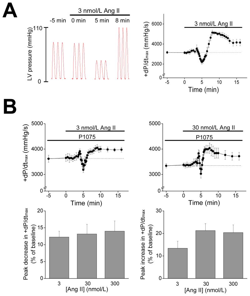
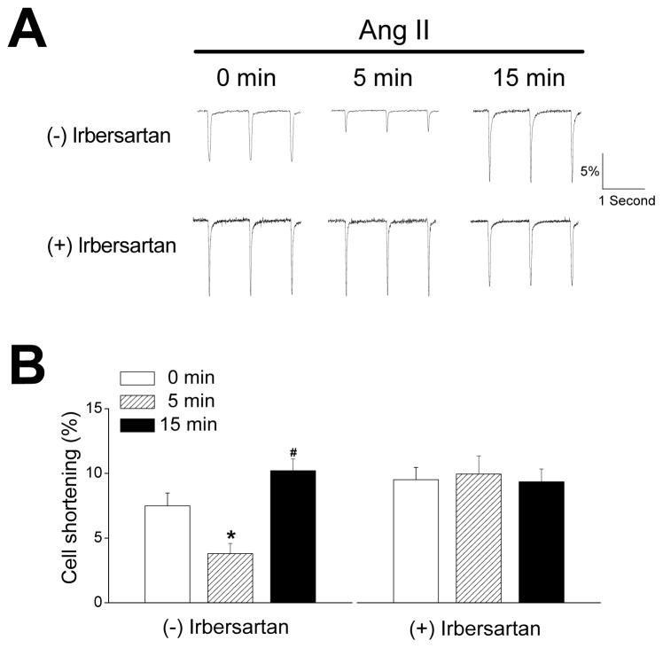
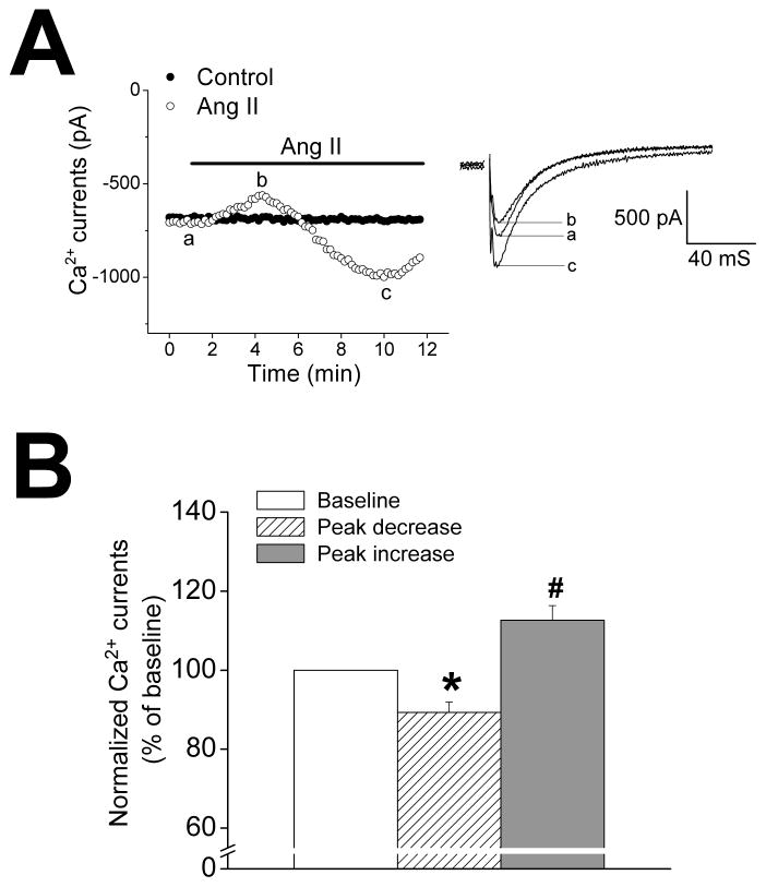
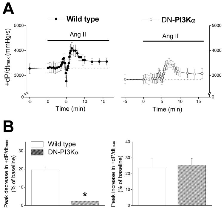
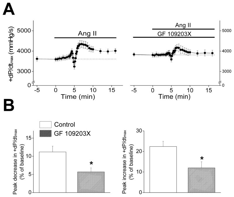
Although angiotensin II (Ang II) plays an important role in heart disease associated with pump dysfunction, its direct effects on cardiac pump function remain controversial. We found that after Ang II infusion, the developed pressure and +dP/dt(max) in isolated Langendorff-perfused mouse hearts showed a complex temporal response, with a rapid transient decrease followed by an increase above baseline. Similar time-dependent changes in cell shortening and L-type Ca(2+) currents were observed in isolated ventricular myocytes. Previous studies have established that Ang II signaling involves phosphoinositide 3-kinases (PI3K). Dominant-negative inhibition of PI3Kalpha in the myocardium selectively eliminated the rapid negative inotropic action of Ang II (inhibited by approximately 90%), whereas the loss of PI3Kgamma had no effect on the response to Ang II. Consistent with a link between PI3Kalpha and protein kinase C (PKC), PKC inhibition (with GF 109203X) reduced the negative inotropic effects of Ang II by approximately 50%. Although PI3Kalpha and PKC activities are associated with glycogen synthase kinase-3beta and NADPH oxidase, genetic ablation of either glycogen synthase kinase-3beta or p47(phox) (an essential subunit of NOX2-NADPH oxidase) had no effect on the inotropic actions of Ang II. Our results establish that Ang II has complex temporal effects on contractility and L-type Ca(2+) channels in normal mouse myocardium, with the negative inotropic effects requiring PI3Kalpha and PKC activities.
Figures
Comment in
-
Negative inotropy by angiotensin II is mediated via phosphoinositide 3-kinase alpha-protein kinase C-coupled signaling pathway.Hypertension. 2010 Sep;56(3):349-50. doi: 10.1161/HYPERTENSIONAHA.110.156158. Epub 2010 Aug 9. Hypertension. 2010. PMID: 20696991 No abstract available.
References
-
- Sadoshima J, Xu Y, Slayter HS, Izumo S. Autocrine release of angiotensin II mediates stretch-induced hypertrophy of cardiac myocytes in vitro. Cell. 1993;75:977–984. - PubMed
-
- Serneri GGN, Boddi M, Cecioni I, Vanni S, Coppo M, Papa ML, Bandinelli B, Bertolozzi I, Polidori G, Toscano T, Maccherini M, Modesti PA. Cardiac angiotensin II formation in the clinical course of heart failure and its relationship with left ventricular function. Circ Res. 2001;88:961–968. - PubMed
-
- Sadoshima J, Izumo S. Molecular characterization of angiotensin II--induced hypertrophy of cardiac myocytes and hyperplasia of cardiac fibroblasts. Critical role of the AT1 receptor subtype. Circ Res. 1993;73:413–423. - PubMed
-
- Gusev K, Domenighetti AA, Delbridge LMD, Pedrazzini T, Niggli E, Egger M. Angiotensin II-mediated adaptive and maladaptive remodeling of cardiomyocyte excitation-contraction coupling. Circ Res. 2009;105:42–50. - PubMed
-
- Moravec C, Schluchter M, Paranandi L, Czerska B, Stewart R, Rosenkranz E, Bond M. Inotropic effects of angiotensin II on human cardiac muscle in vitro. Circulation. 1990;82:1973–1984. - PubMed
Publication types
MeSH terms
Substances
Grants and funding
LinkOut - more resources
Full Text Sources
Miscellaneous
