

doi: 10.1001/archophthalmol.2010.152.
Optic nerve regeneration
Affiliations
- PMID: 20697009
- PMCID: PMC3072887
- DOI: 10.1001/archophthalmol.2010.152
Item in Clipboard
Optic nerve regeneration
Arch Ophthalmol.
2010 Aug.
Abstract
Retinal ganglion cells are usually not able to regenerate their axons after optic nerve injury or degenerative disorders, resulting in lifelong visual loss. This situation can be partially reversed by activating the intrinsic growth state of retinal ganglion cells, maintaining their viability, and counteracting inhibitory signals in the extracellular environment. Advances during the past few years continue to extend the amount of regeneration that can be achieved in animal models. These findings give hope that clinically meaningful regeneration may become a reality within a few years if regenerating axons can be guided to their appropriate destinations.
Figures
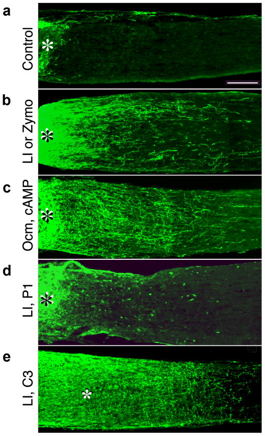
Axon regeneration in the rat optic nerve. Longitudinal sections through the rat optic nerve were stained with antibodies to the protein GAP-43 2 weeks after optic nerve injury to visualize regenerating axons. Asterisks denote the injury site. (a) Almost no regeneration occurs in the absence of further stimulation. (b) Lens injury (LI) or Zy-mosan (Zymo) induces intraocular inflammation and enables RGCs to regenerate axons through the optic nerve, . (c) Ocm plus a cAMP analog, when delivered from slow-release polymeric beads, mimic the effects of lens injury. (d) P1, an Ocm receptor antagonist suppresses the effects of lens injury. (e) Expression of the bacterial enzyme C3 ribosyltransferase (C3) in RGCs blocks the activity of RhoA and enables axons to ignore inhibitory signals in their environment. C3 expression by itself produces only modest levels of regeneration, but greatly enhances the effects of intraocular inflammation after LI.
References
-
- Ramon y Cajal S. Degeneration and Regeneration of the Nervous System. Vol. 5. New York: Oxford University Press; 1991.
-
- Aguayo AJ, Rasminsky M, Bray GM, et al. Degenerative and regenerative responses of injured neurons in the central nervous system of adult mammals. Philos Trans R Soc Lond B Biol Sci. 1991;331:337–343. - PubMed
Publication types
MeSH terms
Substances
Grants and funding
LinkOut - more resources
Full Text Sources
Other Literature Sources
Miscellaneous
