. 2010 Aug;67(8):1002-9.
doi: 10.1001/archneurol.2010.173.

Amyotrophic lateral sclerosis-frontotemporal lobar dementia in 3 families with p.Ala382Thr TARDBP mutations

Affiliations

Amyotrophic lateral sclerosis-frontotemporal lobar dementia in 3 families with p.Ala382Thr TARDBP mutations

Adriano Chiò et al. Arch Neurol. 2010 Aug.

Abstract

Background: TAR DNA-binding protein 43, encoded by the TARDBP gene, has been identified as the major pathological protein of frontotemporal lobar dementia (FTLD) with or without amyotrophic lateral sclerosis (ALS) and sporadic ALS. Subsequently, mutations in the TARDBP gene have been detected in 2% to 3% of patients with ALS (both familial and sporadic ALS). However, to our knowledge, there is only 1 description of 2 patients with FTLD and TARDBP gene mutations who later developed motor neuron disease.

Objective: To describe cognitive abnormalities in 3 Italian families with familial ALS and TARDBP gene mutations.

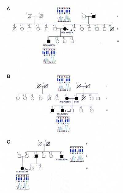
Design, setting, and participants: Genetic, neuropsychological, and neuroimaging analyses in 36 patients with familial non-superoxide dismutase 1 gene (SOD1) ALS and 280 healthy controls. Main Outcome Measure We identified 3 index cases of familial ALS carrying the p.Ala382Thr missense mutation of the TARDBP gene and with clinical, neuroimaging, and neuropsychological features of FTLD.

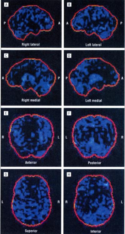
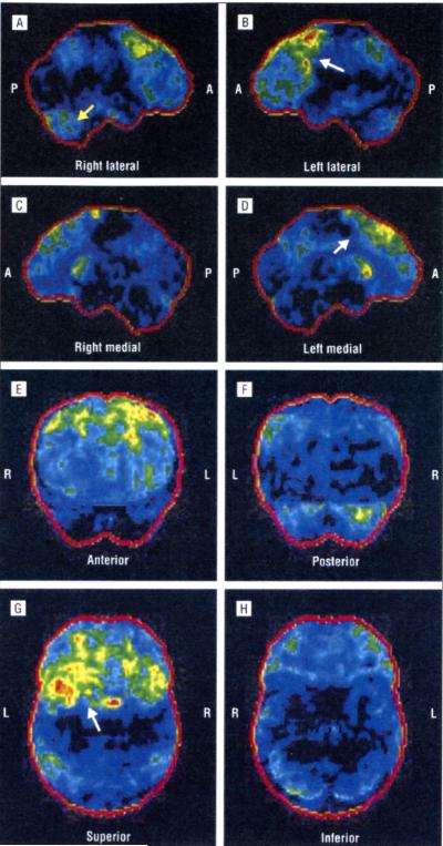
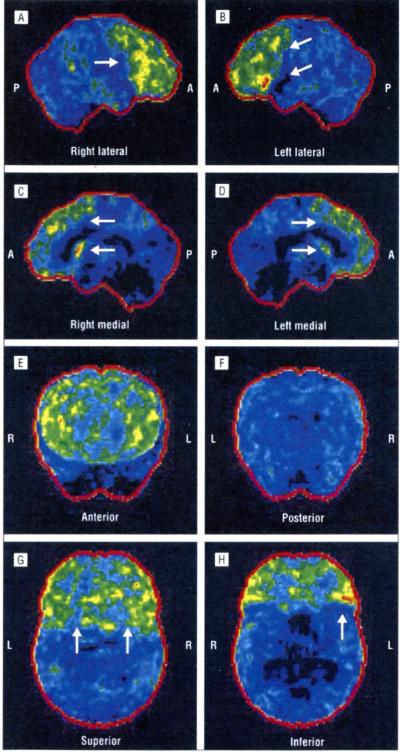
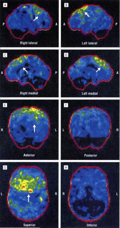
Results: The p.Ala382Thr missense mutation of the TARDBP gene was absent in the 280 controls. It was present in all affected members of the 3 families for whom DNA was available. All affected members of the 3 families developed FTLD after the onset of ALS, confirmed by neuropsychological testing and hypometabolism in frontal associative areas assessed with fludeoxyglucose F 18 positron emission tomography and computed tomography.

Conclusions: Three apparently unrelated families with familial ALS carrying the p.Ala382Thr TARDBP missense mutation developed FTLD. In these families, FTLD cosegregates with ALS. Patients with ALS carrying TARDBP mutations may develop FTLD.


Figures

Figure 1
Figure 1
Pedigrees of family A (A), family B (B), and family C (C) with chromatograms of part of exon 6 of the TARDBP gene showing c.1144G▪A (p.Ala382Thr) mutations in all of the examined patients. Squares indicate males; circles, females; diagonal lines, deceased; open symbols, unaffected; solid symbols, affected; P, proband; and WT, wild type.
Figure 2
Figure 2
Right lateral (A), left lateral (B), right medial (C), left medial (D), anterior (E), posterior (F), superior (G), and inferior (H) positron emission tomography/computed tomography statistical parametric maps of a healthy member of family A. P indicates posterior; A, anterior; R, right; and L, left.
Figure 3
Figure 3
Right lateral (A), left lateral (B), right medial (C), left medial (D), anterior (E), posterior (F), superior (G), and inferior (H) positron emission tomography/computed tomography statistical parametric maps of patient II-7 from family A. The images show the reduction in fludeoxyglucose F 18 uptake on the frontal and parietal associative cortices, the anterior cingulus, and the caudate nuclei. The metabolism is reduced on the left side (white arrows). A slight reduction in fludeoxyglucose F 18 uptake on the right cerebellar cortex (crossed cerebellar diaschisis) is also evident (yellow arrow). P indicates posterior; A, anterior; R, right; and L, left.
Figure 4
Figure 4
Right lateral (A), left lateral (B), right medial (C), left medial (D), anterior (E), posterior (F), superior (G), and inferior (H) positron emission tomography/computed tomography statistical parametric maps of patient III-2 from family B. The images show the hypometabolic areas on the frontal and temporal associative cortices, the frontal mesial cortex, and the caudate nuclei (arrows). P indicates posterior; A, anterior; R, right; and L, left.
Figure 5
Figure 5
Right lateral (A), left lateral (B), right medial (C), left medial (D), anterior (E), posterior (F), superior (G), and inferior (H) positron emission tomography/computed tomography statistical parametric maps of patient III-1 from family C. The images show the hypometabolic areas on the left frontal and temporal associative cortices (arrows). The lesions are less prominent than those of the patients shown in Figure 3 and Figure 4. P indicates posterior; A, anterior; R, right; and L, left.

Similar articles

Cited by

References

    1. Rosen DR, Siddique T, Patterson D, et al. Mutations in Cu/Zn superoxide dismutase gene are associated with familial amyotrophic lateral sclerosis. Nature. 1993;362(6415):5962. - PubMed
    1. Sreedharan J, Blair IP, Tripathi VB, et al. TDP-43 mutations in familial and sporadic amyotrophic lateral sclerosis. Science. 2008;319(5870):16681672. - PMC - PubMed
    1. Kwiatkowski TJ, Jr, Bosco DA, Leclerc AL, et al. Mutations in the FUS/TLS gene on chromosome 16 cause familial amyotrophic lateral sclerosis. Science. 2009;323(5918):12051208. - PubMed
    1. Vance C, Rogelj B, Hortobágyi T, et al. Mutations in FUS, an RNA processing protein, cause familial amyotrophic lateral sclerosis type 6. Science. 2009;323(5918):12081211. - PMC - PubMed
    1. Chow CY, Landers JE, Bergren SK, et al. Deleterious variants of FIG4, a phosphoinositide phosphataseg, in patients with ALS. Am J Hum Genet. 2009;84(1):8588. - PMC - PubMed

Publication types

MeSH terms