. 2010 Oct;186(2):735-55.
doi: 10.1534/genetics.110.119917. Epub 2010 Aug 9.

Refinement of tools for targeted gene expression in Drosophila

Affiliations

Refinement of tools for targeted gene expression in Drosophila

Barret D Pfeiffer et al. Genetics. 2010 Oct.

Abstract

A wide variety of biological experiments rely on the ability to express an exogenous gene in a transgenic animal at a defined level and in a spatially and temporally controlled pattern. We describe major improvements of the methods available for achieving this objective in Drosophila melanogaster. We have systematically varied core promoters, UTRs, operator sequences, and transcriptional activating domains used to direct gene expression with the GAL4, LexA, and Split GAL4 transcription factors and the GAL80 transcriptional repressor. The use of site-specific integration allowed us to make quantitative comparisons between different constructs inserted at the same genomic location. We also characterized a set of PhiC31 integration sites for their ability to support transgene expression of both drivers and responders in the nervous system. The increased strength and reliability of these optimized reagents overcome many of the previous limitations of these methods and will facilitate genetic manipulations of greater complexity and sophistication.


Figures

F<sc>igure</sc> 1.—
Figure 1.—
Diagram of pBP cloning vectors for GAL4, LexA, GAL80, and Split GAL4. All constructs contain the pUC19-derived bacterial origin of replication and ampicillin resistance gene, the PhiC31 attB site, the mini-white marker for identification of transformants in Drosophila, and the DSCP basal promoter (Pfeiffer et al. 2008). The vector backbone is modular to allow for many possible combinations: gray shading indicates components that were held constant, while the colored elements were varied between the constructs we describe in this report. Abbreviations: CRM, conserved regulatory module (generally a 2- to 3-kb enhancer-containing fragment of Drosophila DNA); IVS, intervening sequence within the 5′-UTR; WPRE, a woodchuck hepatitis virus post-transcriptional regulatory element; and TERMINATOR, the transcriptional terminator.
F<sc>igure</sc> 2.—
Figure 2.—
Effects of codon optimization and terminators on GAL4-driven GFP transgene expression. Adult brains are shown after immunostaining to reveal GFP expression. (A) As a control for transcription of the UAS-mCD8∷GFP reporter construct (Lee and Luo 1999) in the absence of a GAL4 driver, as well as for the background of the immunohistochemistry procedure, UAS-mCD8∷GFP was crossed to the attP2 site with no integrated construct. (B–F) The CRM R9C11 fragment (Pfeiffer et al. 2008) was used to drive GAL4 expression in constructs that are integrated into the attP2 site and crossed to the UAS-mCD8∷GFP reporter. (B) The GAL4 gene from Brand et al. (1994), which contains 45 bp of the hsp70 5′-UTR and transcriptional terminators from both the GAL4 gene and the hsp70 gene. CRM R9C11 drives expression prominently in the antennal mechanosensory and motor center (AMMC) and mushroom body (MB). (C) GAL4.1, the same construct as in B, but with a GAL4 coding sequence optimized for Drosophila codon usage. (D) GAL4.2, the same construct as in C, but with the 45-bp hsp70 5′-UTR and yeast transcriptional terminator removed. (E) Same as D, but with GAL4 deletion variant II-9 (pMA236; Ma and Ptashne 1987a) replacing the full-length GAL4 gene. (F) Same as D, but with the SV40 terminator replacing the hsp70 terminator. Relative quantities of GFP mRNA expression levels as measured by QRT–PCR in homogenates of heads of each genotype relative to the control (calibrator) in A, which was arbitrarily set at 1.0, are indicated in the top right of each panel. Assays were done in triplicate except for F, which was done in duplicate; error was within 9% of reported values.
F<sc>igure</sc> 3.—
Figure 3.—
Activation domain choice has a large effect on GAL4-driven levels. Drosophila adult brains were immunostained for GFP after crossing drivers to the UAS-mCD8∷GFP reporter, as in Figure 2. All GAL4 constructs are directed by CRM R9C11, integrated into attP2, and contain an hsp70 terminator. (A) GAL4 from pGawB (Brand and Perrimon 1993). (B) GAL4d, containing the GAL4 deletion variant II-9 (pMA236; Ma and Ptashne 1987a). The 45-bp segment of hsp70 5′-UTR has been removed, but the yeast transcriptional terminator from the GAL4 gene is present. (C) GAL4.2∷VP16, containing a fusion of the GAL4 DNA-binding domain to the VP16 activation domain. The entire coding region has been optimized for Drosophila codon usage and the hsp70 5′-UTR and yeast transcriptional terminator have been removed. (D) GAL4.2∷p65, as in C, but with the activation domain from p65. Relative GFP QRT–PCR values, standardized to attP2 crossed to UAS-mCD8∷GFP (set at 1.0), are indicated in the top right of each panel. Assays were done in triplicate; error was within 5% of reported values.
F<sc>igure</sc> 4.—
Figure 4.—
Diagram of pJFRC reporter constructs. All constructs contain the pUC19-derived bacterial origin of replication and ampicillin resistance gene, the PhiC31 attB site, the mini-white marker for identification of transformants in Drosophila, an hsp70 basal promoter, and an SV40 transcriptional terminator. The vector backbone is modular to allow for many possible combinations: gray shading indicates components that were held constant, while the colored elements were varied between the constructs we describe in this report. Examples of some of these alternatives are listed below colored elements; see text for more details.
F<sc>igure</sc> 5.—
Figure 5.—
Increasing the number of GAL4 DNA-binding sites boosts GFP levels. Drosophila adult brains were immunostained for GFP after crossing R9C11-GAL4 to mCD8∷GFP reporters with 5 to 40 UAS sites. With the exception of the UAS-mCD8∷GFP construct of Lee and Luo (1999), which is a P-element insertion on the second chromosome, all constructs are integrated into attP2. (A) attP2 (no GAL4 driver) crossed to pJFRC2 (negative control). (B–F) R9C11-GAL4 crossed to reporters containing (B) 5, (C) 10, (D) 15, (E) 20, and (F) 40 UAS sites. We also tested a 5XUAS construct in the pJFRC backbone (pJFRC5; see Table 2), which gave similar results to UAS-mCD8∷GFP (data not shown). Relative GFP QRT–PCR values, normalized to a level of 1.0 for attP2 × UAS-mCD8∷GFP (see Figure 2A), are indicated in the top right of each panel. Assays were done in triplicate; error was within 7% of reported values.
F<sc>igure</sc> 6.—
Figure 6.—
Tandem reporters can be used to increase output. (A) Diagram of reporter constructs. Note variation in UAS copy number (indicated in the blue box), tandem orientation (indicated by the arrows), and presence of a gypsy-insulated spacer of 2.8 kb (INS). The synthesized spacer was designed by independently randomizing a minimum of five times (Shuffle program GCG Version 11.1; Accelrys, San Diego) a sequence derived from kanamycin CDS (base pairs 1–810) and a sequence derived from the E. coli lacZ CDS (base pairs 799–2000) and then fusing these randomized sequences. The spacer was then flanked on either end with 424 bp from the 5′-UTR of gypsy (base pairs 647–1074) that contains 12 binding sites for the su(Hw) protein (Marlor et al. 1986; Spana et al. 1988). (B–F) Drosophila adult brains immunostained for GFP after crossing R9C11-GAL4 driver to indicated reporter. All constructs integrated into attP2. (B and C) Controls showing driver pattern with (B) 10 UAS copies (pJFRC2) or (C) 20 copies (pJFRC7). (D) A tandem 10XUAS-mCD8∷GFP reporter in a tail-to-head orientation (pJFRC9). (E) As in D, but with inclusion of a gypsy-insulated 2.8-kb spacer between inserts (pJFRC10). (F) As in E, but reporters are inserted tail to tail (pJFRC11). Relative GFP QRT–PCR values, standardized to attP2 × UAS-mCD8∷GFP, are indicated in the top right corners. Assays were done in duplicate; error was within 13% of reported values.
F<sc>igure</sc> 7.—
Figure 7.—
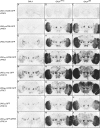
Tuning expression levels by varying the strength of the activation domain and UAS responder. Drosophila adult brains were immunostained for GFP. With the exception of the 5× UAS-mCD8∷GFP (Lee and Luo 1999) construct all transgenes are integrated into attP2. CRM R9B05 was used to drive three GAL4 variants: standard GAL4 (as used in the constructs described by Pfeiffer et al. 2008), GAL4.2∷VP16, or GAL4.2∷p65. These three GAL4 drivers were crossed to different responders as indicated, which vary in number of UAS sites, localization tag, and inclusion of a WPRE: (A–C) The 1XUAS-mCD8∷GFP (pJFRC3). (D–F) The 3XUAS-mCD8∷GFP (pJFRC4). (G–I) The 5XUAS-mCD8∷GFP of Lee and Luo (1999). (J–L) The 10XUAS-mCD8∷GFP (pJFRC2). (M–O) The 10XUAS-myr∷GFP (pJFRC12): myristoylated, codon-optimized GFP. (P–R) The 20XUAS-mCD8∷GFP (pJFRC7). (S–U) The 10XUAS-GFP (pJFRC13): untagged (cytoplasmic), codon-optimized GFP. (V–X) As in S–U, but containing a WPRE in the 3′-UTR. Note that strength of the activation domain and UAS number can drive undesirable levels of GFP and cause cytotoxicity. (P and R) Weakly expressing cells in the optic lobe become prominent, while those that expressed robust levels are gone (see Figure S2).
F<sc>igure</sc> 8.—
Figure 8.—
Improved LexA drivers with GADfl and p65 activation domains. Drosophila adult brains were immunostained for GFP after crossing LexA drivers to a published LexAop-rCD2∷GFP (Lai and Lee 2006). All GAL4 and LexA constructs are directed by CRM R9C11 and integrated into attP2. (A) attP2 (no LexA driver) crossed to published LexAop-rCD2∷GFP (negative control; note “leak” expression in the lamina). (B) R9C11-GAL4 with UAS-mCD8∷GFP. (C and D) R9C11-LexA drivers containing GAL4 activation domain variants GADd (C) or GADfl (D), crossed to rCD2∷GFP. (E) As in D, but with a nuclear localization signal (nls). (F and G) R9C11-LexA drivers with GAL80-insensitive activation domains VP16 (F) or p65 (G), crossed to rCD2∷GFP. (H) As in G, but with an nls. Relative GFP mRNA levels as measured by QRT–PCR, standardized to attP2 × UAS-mCD8∷GFP (set as 1.0), are indicated in the top right of each panel. Assays were done in duplicate; error was within 14% of reported values.
F<sc>igure</sc> 9.—
Figure 9.—
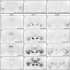
A LexA operator containing 13 binding sites from sulA provides robust and nonleaky expression. Drosophila adult brains were immunostained for GFP. With the exception of LexAop-rCD2∷GFP (Lai and Lee 2006) all constructs are integrated into attP2. (A) attP2 (no LexA driver) crossed to LexAop-rCD2∷GFP (negative control; note “leak” expression in the lamina indicated by arrowheads). (B) attP2 (no LexA driver) crossed to pJFRC15-13XLexAop2-mCD8∷GFP (pJFRC15; negative control, no detectable leak), a LexAop reporter containing 13 LexA-binding sites derived from sulA. (C) attP2 (no GAL4 driver) crossed to pJFRC2 (negative control). (D–L) Enhancers driving LexA variants (indicated in the bottom right of each panel) crossed to the LexA reporter pJFRC15: (D, G, and J) R9D11-LexA; (E, H, and K) R9C11-LexA; (F, I, and L) R9B05-LexA. (M–O) GAL4 driven by the same three enhancers crossed to pJFRC2: (M) R9D11-GAL4, (N) R9C11-GAL4, and (O) R9B05-GAL4.
F<sc>igure</sc> 10.—
Figure 10.—
Refinement of GFP expression using Split GAL4. Drosophila third instar central nervous systems were immunostained for GFP. All constructs are integrated into attP2. (A) attP2 (no GAL4 driver) crossed to pJFRC2-10XUAS-IVS-mCD8∷GFP (negative control). (B and C) R20B05p65AD or R35B08GAL4DBD with pJFRC2 (negative control: no detectable leak). (D) R20B05-GAL4 crossed to pJFRC2 drives expression in immature neurons of the optic lobes and in all of the lineages of secondary neurons in the central brain and ventral nerve cord but none of the primary neurons that are born during embryogenesis. (E) R35B08 crossed to pJFRC2 drives expression in primary neurons of the central brain and ventral nerve cord, as well as eight secondary lineages in the central brain and secondary lineages 0, 8, and 7 in the thoracic neuromeres of the ventral nerve cord (for a description of lineages see Truman et al. 2004). (F) Crossing Split GAL4 DNA-binding domain driven by R35B08 (R35B08GAL4DBD; enhancer fragment R35B08 cloned in vector pBPZpGAL4DBDUw) with a stock of R20B05p65AD (enhancer fragment R20B05 cloned in vector pBPp65ADZpUw); pJFRC2 (attP40; attP2) yields GFP expression restricted to the overlap between the two parent patterns. (G) R50B06 crossed to pJFRC2 drives expression in primary neurons of the brain and central nervous system, secondary lineage BAmv3 (for a description of brain lineages, see Pereanu and Hartenstein 2006) in the central brain, and secondary lineages 9, 17, and 23 of the ventral nerve cord. (H) Crossing Split GAL4 DNA-binding domain driven by R50B06 (R50B06GAL4DBD) with a stock of R20B05p65AD; pJFRC2 (attP40; attP2) yields GFP expression restricted to the overlap between the two parent patterns. In both F and H, expression in primary neurons is eliminated, as predicted.
F<sc>igure</sc> 11.—
Figure 11.—
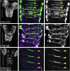
Targeted refinement of a GAL4 pattern using GAL80. Dissected third instar central nervous systems were immunostained for GFP (green) and neurotactin (magenta). All GAL4 and GAL80 constructs are integrated in attP2; the reporter is a P-element insertion of UAS-mCD8∷GFP on the second chromosome (Lee and Luo 1999). (A) R20B05 drives expression in the immature neurons of the optic lobes (OL) and in all of the lineages of secondary neurons in the central brain and ventral nerve cord in third instar larvae. (B) A single optical section of the region indicated by the blue line in A at the level of the intermediate commissures. R20B05 drives GFP expression in both anterior and posterior commissures of thoracic segments T1–T3 and abdominal segment A1: anterior commissures are indicated by solid yellow arrowheads. (C) As in B, but only the green channel (anti-GFP) is shown. (D) R15E07 drives expression in most of the secondary lineages in the central brain and VNC, with the notable exception of the neuroblast lineages whose axon bundles comprise the anterior intermediate commissure in the thoracic segments. It does not drive optic lobe expression. (E) A single optical section of the region indicated by the blue line in D. Note that R15E07 drives GFP expression in only the posterior commissures; the anterior commissures lacking GFP expression are indicated by open yellow arrowheads. (F) As in E, but only the green channel (anti-GFP) is shown. (G) R15E07-GAL80-SV40 (attP40) crossed to w; UAS-mCD8∷GFP; R20B05-GAL4. As predicted, GFP expression is now restricted to the optic lobes (OL), a few lineages in the central brain, and the thoracic and abdominal lineages that form the anterior intermediate commissure. (H) A single optical section of the region indicated by the blue line in G showing GFP expression restricted to the lineages of the anterior commissure: anterior commissures are indicated by solid yellow arrowheads. (I) As in H, but only the green channel (anti-GFP) is shown.
F<sc>igure</sc> 12.—
Figure 12.—
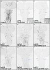
GAL80 suppression of GAL4 is improved with post-transcriptional regulatory elements (intron and WPRE). Drosophila third instar central nervous systems were immunostained for GFP. Enhancer R11F05 was used to drive different GAL80 constructs. These constructs were then tested against their parent enhancer using a recombinant reporter line of R11F05-GAL4 (attP2) and a P-element insertion of UAS-mCD8∷GFP on the third chromosome. Both GAL4 and GAL80 constructs use an hsp70 terminator, unless otherwise noted. GFP expression was assayed after crossing R11F05-GAL4 (attP2) UAS-mCD8∷GFP to (A) Canton S (positive control), showing GFP expression in a subset of sensory neurons that project into the ventral nerve cord, and (B) R11F05-GAL80-SV40 (no post-transcriptional regulatory elements), integrated in attP2. The inset at the bottom right shows a portion of the VNC at higher gain. (C) As in B, but integrated in attP40. The inset at the bottom right shows a portion of the VNC at higher gain. (D) R11F05-GAL80, integrated into attP2. A small number of neurons in the pattern escape suppression (express GFP). (E) As in D, but integrated into the weaker site attP40. Note increased incidence of neurons that escape suppression. (F–I) Inclusion of post-transcriptional regulatory elements to R11F05-GAL80 increases the level of GFP suppression: (F) R11F05-GAL80 with a WPRE, in attP40; (G) R11F05-GAL80 with an IVS, in attP40; (H) R11F05-GAL80 with both IVS and WPRE, in attP40; (I) R11F05-GAL80 with both IVS and WPRE, in attP2. Weak background GFP expression from GAL4-independent expression from the UAS-mCD8∷GFP reporter was present in a small group of cells in all preparations (examples indicated by arrowheads).
F<sc>igure</sc> 13.—
Figure 13.—
Map of evaluated genomic attP sites. The indicated 16 PhiC31 genomic attP integration sites were assayed for four properties: (1) expression in the adult nervous system when an enhancer trap vector was inserted, (2) expression from an exogenous enhancer, (3) expression from a UAS construct responding to a GAL4 driver, and (4) transgene integration rate. Sites shown in red meet all four criteria, while those shown in blue performed well with UAS reporter constructs, but not with enhancer-GAL4 drivers. Sites shown in black were rejected. See text for details. Genomic attP site references: attP1, attP2, attP3, attP18, and attP40 (Groth et al. 2004; Markstein et al. 2008); attP16 (Markstein et al. 2008); VK00005, VK00016, VK00026, and VK00027 (Venken et al. 2006); and su(Hw)attP1, su(Hw)attP2, su(Hw)attP4, su(Hw)attP5, su(Hw)attP6, and su(Hw)attP8 (Ni et al. 2009; this study).
F<sc>igure</sc> 14.—
Figure 14.—
Chromatin effects on R9C11-GAL4. Drosophila adult brains were immunostained for GFP. R9C11-GAL4 was integrated into 10 attP docking sites and crossed to pJFRC2 in attP2. The genomic docking site is shown in the top left corner of each panel and the chromosome arm of the insertion site in the top right. We were unable to get transformants in the attP3 and VK00026 sites. Four sites showed reproducible and robust expression, comparable to our standard attP2 insertion: attP18, su(Hw)attP8, attP40, and VK00027. Genomic docking sites flanked with gypsy insulators [indicated by su(Hw) in the name] share a common background leak in a half-dozen cells in the lateral horn (see Figure S3 and text).
F<sc>igure</sc> 15.—
Figure 15.—
Chromatin effects on pJFRC2, assayed by R9C11-GAL4. Drosophila adult brains were immunostained for GFP. pJFRC2 was integrated in 12 docking sites and crossed to R9C11-GAL4 in attP2. The genomic docking site is shown in the top left corner of each panel and the chromosome arm of the insertion site in the top right. With the exception of VK00026, all docking sites showed strong, reproducible expression (also see Figure S6 and Figure S7). UAS transgenes seem to be less susceptible to chromatin influence than enhancer-GAL4 constructs; for example, VK00005 works well for UAS, but not GAL4, insertions (compare with Figure 14; also see Figure S4 and Figure S5).

Similar articles

Cited by

References

    1. Adams, M. D., S. E. Celniker, R. A. Holt, C. A. Evans, J. D. Gocayne et al., 2000. The genome sequence of Drosophila melanogaster. Science 287 2185–2195. - PubMed
    1. Ahmad, K., and S. Henikoff, 2001. Modulation of a transcription factor counteracts heterochromatic gene silencing in Drosophila. Cell 104 839–847. - PubMed
    1. Atasoy, D., Y. Aponte, H. H. Su and S. M. Sternson, 2008. A FLEX switch targets Channelrhodopsin-2 to multiple cell types for imaging and long-range circuit mapping. J. Neurosci. 28 7025–7030. - PMC - PubMed
    1. Bellen, H. J., C. Wilson and W. J. Gehring, 1990. Dissecting the complexity of the nervous system by enhancer detection. BioEssays 12 199–204. - PubMed
    1. Bischof, J., R. K. Maeda, M. Hediger, F. Karch and K. Basler, 2007. An optimized transgenesis system for Drosophila using germ-line-specific phiC31 integrases. Proc. Natl. Acad. Sci. USA 104 3312–3317. - PMC - PubMed

Publication types

MeSH terms

LinkOut - more resources