. 2010 Oct 21;29(42):5741-54.
doi: 10.1038/onc.2010.309. Epub 2010 Aug 9.

MUC4 mucin-induced epithelial to mesenchymal transition: a novel mechanism for metastasis of human ovarian cancer cells

Affiliations

MUC4 mucin-induced epithelial to mesenchymal transition: a novel mechanism for metastasis of human ovarian cancer cells

M P Ponnusamy et al. Oncogene. .

Erratum in

  • Oncogene. 2010 Nov 11;29(45):6084

Abstract

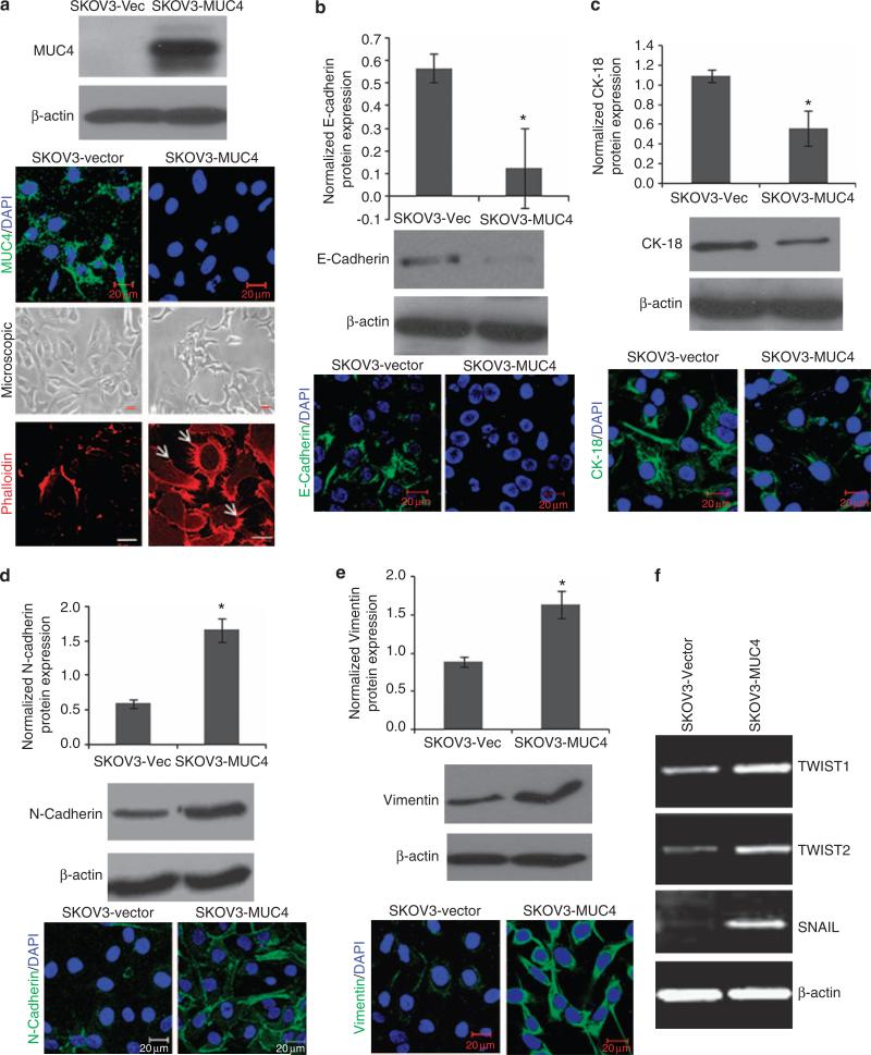
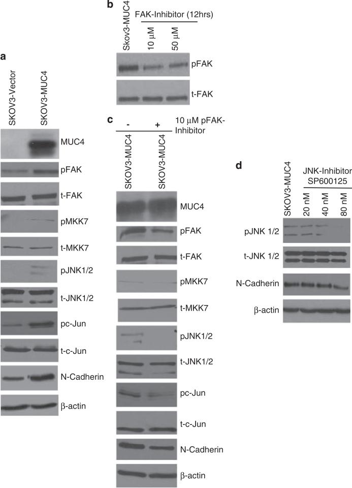
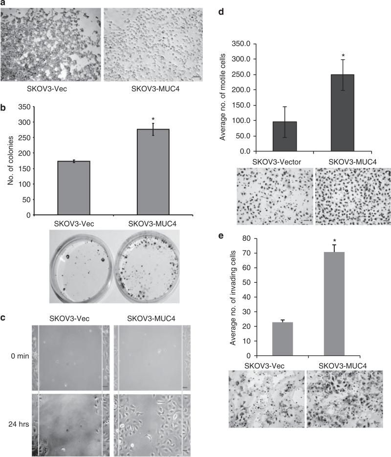
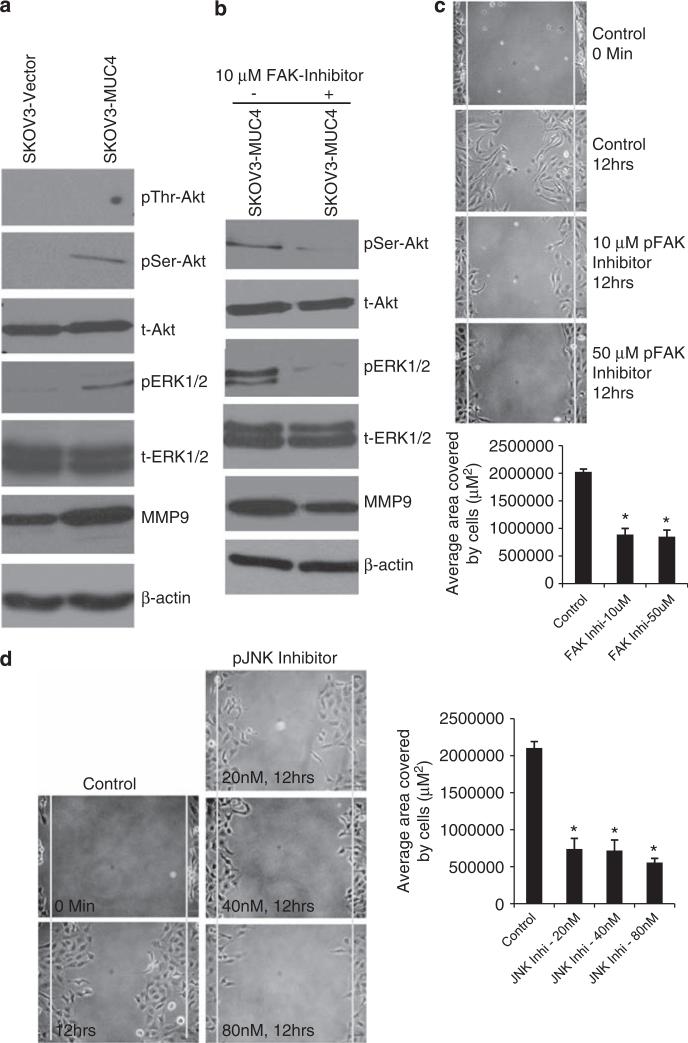
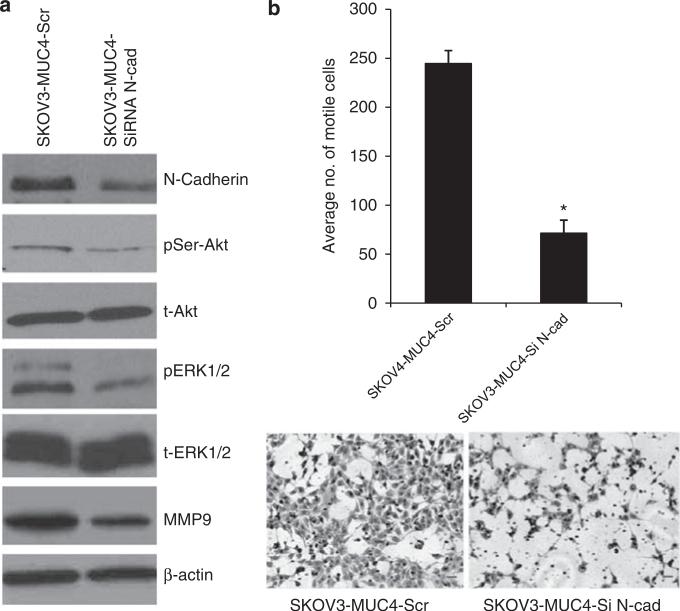
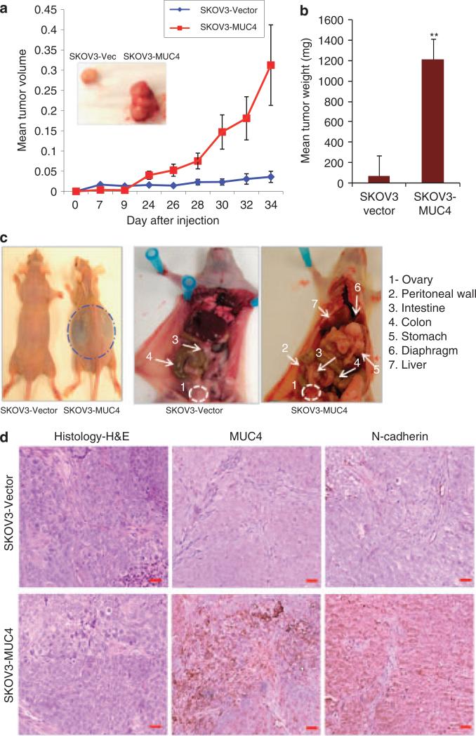
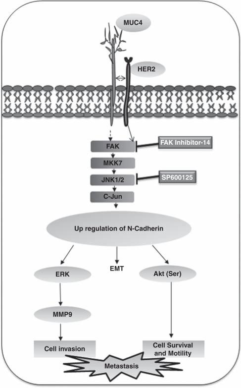
The acquisition of invasiveness in ovarian cancer (OC) is accompanied by the process of epithelial-to-mesenchymal transition (EMT). The MUC4 mucin is overexpressed in ovarian tumors and has a role in the invasiveness of OC cells. The present study was aimed at evaluating the potential involvement of MUC4 in the metastasis of OC cells by inducing EMT. Ectopic overexpression of MUC4 in OC cells (SKOV3-MUC4) resulted in morphological alterations along with a decreased expression of epithelial markers (E-cadherin and cytokeratin (CK)-18) and an increased expression of mesenchymal markers (N-cadherin and vimentin) compared with the control cells (SKOV3-vector). Also, pro-EMT transcription factors TWIST1, TWIST2 and SNAIL showed an upregulation in SKOV3-MUC4 cells. We further investigated the pathways upstream of N-cadherin, such as focal adhesion kinase (FAK), MKK7, JNK1/2 and c-Jun, which were also activated in the SKOV3-MUC4 cells compared with SKOV3-vector cells. Inhibition of phospho-FAK (pFAK) and pJNK1/2 decreased N-cadherin expression in the MUC4-overexpressing cells, which further led to a significant decrease in cellular motility. Knockdown of N-cadherin decreased the activation of extracellular signal-regulated kinase-1/2 (ERK1/2), AKT and matrix metalloproteinase 9 (MMP9), and inhibited the motility in the SKOV3-MUC4 cells. Upon in vivo tumorigenesis and metastasis analysis, the SKOV3-MUC4 cells produced significantly larger tumors and demonstrated a higher incidence of metastasis to distance organs (peritoneal wall, colon, intestine, stomach, lymph nodes, liver and diaphragm). Taken together, our study reveals a novel role for MUC4 in inducing EMT through the upregulation of N-cadherin and promoting metastasis of OC cells.


Figures

Figure 1
Figure 1
MUC4 overexpression alters cell morphology and epithelial phenotype marker expression. (a) Immunoblotting analysis of ectopic MUC4 expression in SKOV3 cells (SKOV3-MUC4) and control SKOV3 cells (SKOV3-vector). First panel showed the confocal immunofluorescence analysis for MUC4 in SKOV3-vector and SKOV3-MUC4 cells (scale bar-20 μm; fluorescein isothiocyanate (FITC) for MUC4 and 4′-6-diamidino-2-phenylindole (DAPI) for nuclear staining). Middle panel showed the phase contrast microscopic picture of MUC4-overexpressing SKOV3 cells and control cells (original magnification × 200, scale bar-0.8 mm). The last panel showed the phalloidin-rhodamine (phalloidin-RITC) staining of actin–cytoskeleton variation in both empty vector- and MUC4-transfected SKOV3 cells (scale bar-20 μm). MUC4-overexpressed SKOV3 cells were associated with the presence of more microspikes, lamellopodia- and filopodia-like cellular projections (arrows) with dense actin concentrated at the cellular protrusions compared with the empty vector control cells. (b, c) Immunoblotting analysis showed significantly decreased expression of epithelial markers E-cadherin (*P = 0.021) and CK-18 (*P = 0.01) in SKOV3-MUC4 cells compared with SKOV3-vector cells. Confocal microscopy showed increased staining of E-cadherin and CK in the SKOV3-vector cells and diminished staining in SKOV3-MUC4 cells (scale bar-20 μm). (FITC-conjugated goat anti-mouse IgG for secondary antibody and DAPI was used for nuclear staining.) (d, e) Immunoblotting analysis showed the significantly increased expression of mesenchymal marker N-cadherin (*P = 0.017) and vimentin (*P = 0.04) in SKOV3-MUC4 cells compared with SKOV3-vector cells. Confocal microscopy showed increased staining of N-cadherin and vimentin in the SKOV3-MUC4 cells and decreased staining in SKOV3-vector cells (scale bar-20 μm). (FITC-conjugated goat anti-mouse IgG for secondary antibody and DAPI was used for nuclear staining.) (f) Reverse transcriptase–PCR analysis for the EMT key transcription factors TWIST1, TWIST2 and SNAIL in both SKOV3-MUC4 and SKOV3-vector cells. β-actin was used as a control.
Figure 2
Figure 2
MUC4 expression induces upregulation of N-cadherin via FAK signaling. (a) Immunoblot analysis showed an increase in the activation of FAK, MKK7, JNK1/2, c-Jun and upregulate N-cadherin in SKOV3-MUC4 cells as compared with vector control cells. The total form of FAK, MKK7, JNK1/2, c-Jun molecules remains unchanged in both cell lines. β-actin was used as a loading control. (b) Treatment with pFAK inhibitor: Immunobloting analysis of pFAK and FAK in cells treated with 10 and 50 μm FAK inhibitor (FAK inhibitor-14 (1,2,4,5-benzenetetraamine tetrahydrochloride)-treated SKOV3-MUC4 cells. (c) Western blot analysis showed that the inhibition of pFAK reduces the activation of FAK, MKK7, JNK1/2, c-Jun and decreases the expression of N-cadherin. The total form of FAK, MKK7, JNK1/2, c-Jun molecules remains the same. β-actin was used as a loading control. (d) Inhibition of pJNK1/2: immunoblotting analysis showing the expression of pJNK, JNK and N-cadherin in JNK1/2 inhibitor (SP600125; 20, 40 and 80 nm)-treated SKOV3-MUC4 cells. β-actin was used as a loading control.
Figure 3
Figure 3
MUC4 decreases the aggregation and increases the colonogenecity, motility and invasiveness of SKOV3 cells. (a) Aggregation assay: a reduced cellular aggregation was observed in MUC4-overexpressing SKOV3 cells, whereas vector-transfected cells (SKOV3-vector) showed bigger and tight cell aggregates (original magnification × 100, scale bar-0.8 mm). (b) Colony forming assay: MUC4-transfected SKOV3 cells formed a significantly greater number of (*P = 0.029) colonies than the control cells (original magnification × 40, scale bar-0.8 mm). (c) Wound healing assay was performed to visualize the differences in motility of MUC4- and vector-transfected SKOV3 cell lines (original magnification × 100, scale bar-0.8 mm). (d) Boyden's chamber motility assay for both MUC4- and empty vector-transfected SKOV3 cells. Cell motility was observed to be significantly (*P = 0.0001) increased in MUC4-transfected SKOV3 cells (original magnification × 100, scale bar-0.8 mm). (e) Matrigel-coated Boyden's chamber invasion assay. SKOV3-MUC4 cells exhibited a significant increase in invasiveness compared with the SKOV3-vector cells (*P = 0.00055; original magnification × 100, scale bar-0.8 mm).
Figure 4
Figure 4
Analysis of N-cadherin downstream signaling pathways in MUC4-expressing and control cells. (a) Western blot analysis of N-cadherin downstream signaling molecules showed increased activation of serine (473) Akt, ERK1/2 and increased expression of MMP9 in MUC4-overexpressed cells. No variation was seen in threonine (Y308) phosphorylated Akt. The total form of Akt and ERK1/2 were unchanged in both the SKOV3-derived sublines. β-actin was used as a loading control. (b) Western blot analysis showed that pFAK inhibition also reduces the phosphorylation of serine (473) Akt, ERK1/2 and decreases the expression of MMP9 compared with the mock control. (c, d) Wound healing assay: The scratch was made across the cell monolayer in pFAK (1,2,4,5-benzenetetraamine tetrahydrochloride) and pJNK (SP600125) inhibitor-treated SKOV3-MUC4 cells with mock control. The migration of cells was measured (μm2) in treated and untreated cells. Significant migration of cells was analyzed using two-tailed Student's t-test. A P-value of <0.05 was considered statistically significant. (FAK Inhi-10 mm, *P <0.0004; FAK Inhi-50 μm, *P <0.0003), (JNK1/2 Inhi-20 nm, *P <0.005; JNK1/2 Inhi-40 nm, *P <0.0002; JNK1/2 Inhi-80 nm, *P <0.0001) (original magnification × 100, scale bar-0.8 mm).
Figure 5
Figure 5
Knockdown of N-cadherin in MUC4-overexpressing cells reduces their motility. (a) The transient knockdown of N-cadherin using three pooled siRNA oligos for 48 h in SKOV3-MUC4 cells. Western blot analysis showed a downregulation of N-cadherin in siRNA-treated SKOV3-MUC4 cells compared with scramble RNA interference (RNAi)-treated SKOV3-MUC4 cells. The phosphorylated (Ser) Akt and pERK1/2 were reduced in N-cadherin knockdown cells compared with control cells. The total form of Akt and ERK, however, remained unchanged. MMP9 expression was also reduced in the N-cadherin knockdown cells. β-actin was used as a loading control. (b) Motility assay in N-cadherin knockdown SKOV3-MUC4 cells. SKOV3-MUC4 cells were treated with N-cadherin siRNA and after 24 h were trypsinized for the motility assay as described previously. There was a significant decrease in motility in the N-cadherin siRNA-treated cells compared with the scramble RNAi-treated cells (*P = 0.004; original magnification × 100, scale bar-0.8 mm).
Figure 6
Figure 6
Analysis of tumor growth and metastasis in the MUC4-transfected OC cells. (a, b) In vivo tumorigenesis assay for SKOV3-MUC4 and SKOV3-vector cells with NUDE/SCID mice following subcutaneous implantation. Measurement of tumor volumes indicated that SKOV3-MUC4 cells formed significantly larger tumors as compared with control cells. Inset shows a representative image for tumors developed in SKOV3-vector- and SKOV3-MUC4-injected mice. Tumor weight was significantly higher (**P = 0.00001) in the SKOV3-MUC4 cells compared with SKOV3-vector cells. (c) In vivo metastasis analysis for SKOV3-MUC4 and SKOV3-vector cells with NUDE/SCID mice. The SKOV3-MUC4 cells injected mice showed more ascitic fluid accumulation. The ovary was found to develop tumors in both groups of mice. The SKOV3-vector-injected mice showed fewer incidence of metastasis in distant organ sites, whereas SKOV3-MUC4-injected mice showed metastatic deposits in the peritoneal wall, small intestine, colon, stomach, liver and diaphragm. (d) Hematoxylin and eosin staining of tumors produced by i.p. injection of SKOV3-vector and SKOV3-MUC4 cells. The immunohistochemical analysis of MUC4 and N-cadherin showed increased reactivity in the SKOV3-MUC4 tumors compared with SKOV3-vector tumors (original magnification × 200, scale bar-0.8 mm).
Figure 7
Figure 7
Schematic representation of the proposed mechanism of MUC4-induced EMT and metastasis of OC. MUC4 induces the FAK activation either directly (unknown mechanism) or by interacting with HER2 cause the activation of MKK7, JNK1/2 and c-Jun and leading to upregulation of N-cadherin. The upregulation of N-cadherin induces the EMT process in OC cells. Furthermore, the N-cadherin downstream signaling of Akt, ERK1/2 and MMP9 may be the primary cause for an increased motility and invasion in MUC4-expressing OC cells.

Similar articles

Cited by

References

    1. Ahmed N, Thompson EW, Quinn MA. Epithelial-mesenchymal interconversions in normal ovarian surface epithelium and ovarian carcinomas: an exception to the norm. J Cell Physiol. 2007;213:581–588. - PubMed
    1. Andrianifahanana M, Moniaux N, Schmied BM, Ringel J, Friess H, Hollingsworth MA, et al. Mucin (MUC) gene expression in human pancreatic adenocarcinoma and chronic pancreatitis: a potential role of MUC4 as a tumor marker of diagnostic significance. Clin Cancer Res. 2001;7:4033–4040. - PubMed
    1. Auersperg N, Pan J, Grove BD, Peterson T, Fisher J, Maines-Bandiera S, et al. E-cadherin induces mesenchymal-to-epithelial transition in human ovarian surface epithelium. Proc Natl Acad Sci USA. 1999;96:6249–6254. - PMC - PubMed
    1. Behrens J. Cadherins as determinants of tissue morphology and suppressors of invasion. Acta Anat (Basel) 1994;149:165–169. - PubMed
    1. Boman F, Buisine MP, Wacrenier A, Querleu D, Aubert JP, Porchet N. Mucin gene transcripts in benign and borderline mucinous tumours of the ovary: an in situ hybridization study. J Pathol. 2001;193:339–344. - PubMed

Publication types

MeSH terms