. 2010 Oct 21;29(42):5667-77.
doi: 10.1038/onc.2010.327. Epub 2010 Aug 9.

The reactive tumor microenvironment: MUC1 signaling directly reprograms transcription of CTGF

Affiliations

The reactive tumor microenvironment: MUC1 signaling directly reprograms transcription of CTGF

M E Behrens et al. Oncogene. .

Abstract

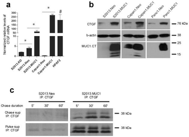
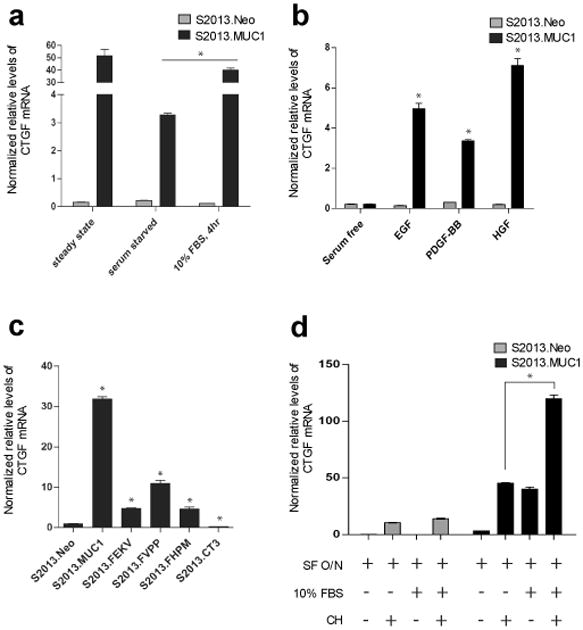
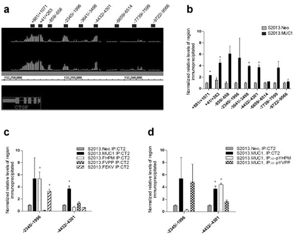
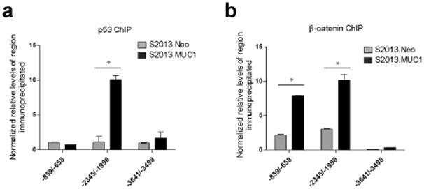
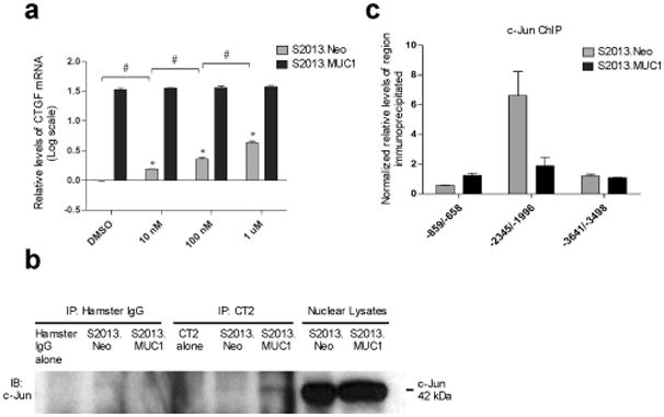
The MUC1 cytoplasmic tail (MUC1.CT) conducts signals from spatial and extracellular cues (growth factor and cytokine stimulation) to evoke a reprogramming of the cellular transcriptional profile. Specific phosphorylated forms of the MUC1.CT achieve this function by differentially associating with transcription factors and redirecting their transcriptional regulatory capabilities at specific gene regulatory elements. The specificity of interaction between MUC1.CT and several transcription factors is dictated by the phosphorylation pattern of the 18 potential phosphorylation motifs within the MUC1.CT. To better appreciate the scope of differential gene expression triggered by MUC1.CT activation, we performed microarray gene expression analysis and chromatin immunoprecipitation (ChIP)-chip promoter analysis and identified the genome-wide transcriptional targets of MUC1.CT signaling in pancreatic cancer. On a global scale, MUC1.CT preferentially targets genes related to invasion, angiogenesis and metastasis, suggesting that MUC1.CT signaling contributes to establishing a reactive tumor microenvironment during tumor progression to metastatic disease. We examined in detail the molecular mechanisms of MUC1.CT signaling that induces the expression of connective tissue growth factor (CTGF/CCN2), a potent mediator of ECM remodeling and angiogenesis. We demonstrate a robust induction of CTGF synthesis and secretion in response to serum factors that is enabled only when MUC1 is highly expressed. We demonstrate the requirement of phosphorylation at distinct tyrosine motifs within the MUC1.CT for MUC1-induced CTGF expression and demonstrate a phosphorylation-specific localization of MUC1.CT to the CTGF promoter. We found that MUC1 reorganizes transcription factor occupancy of genomic regions upstream of the CTGF gene, directing β-catenin and mutant p53 to CTGF gene regulatory elements to promote CTGF expression and destabilizing the interaction at these regions of the transcriptional repressor, c-Jun. With this example we illustrate the capacity of MUC1.CT to mediate transcription factor activity in a context-dependent manner to achieve wide spread and robust changes in gene expression and facilitate creation of the reactive tumor microenvironment.


Conflict of interest statement

Confilct of interest: The authors declare no conflict of interest

Figures

Figure 1
Figure 1
Overexpression of MUC1 increases CTGF levels in pancreatic cancer cell lines. (a) MUC1 overexpression increases CTGF mRNA transcripts in S2013 and Capan1 cells. Stable knockdown of MUC1 in S2013 cells further reduces CTGF transcripts when compared to S2013.Neo. Elevated relative levels of CTGF mRNA levels are observed in HPAF2 cells (#, P=0.094 when compared to S2013.Neo). All values have been normalized to levels of β-actin and are expressed as the average of three reactions +/− standard error mean (*, P<0.05). (b) Elevated levels of CTGF are observed at the protein level with MUC1 overexpression in pancreatic cancer cell lines S2013, Capan1, and Panc1. (c) Following metabolic radiolabeling using 35S-containing culture media for 1 hour (Pulse), media was collected (Pulse supe) and replaced with “cold” Cysteine and Methionine-containing media (Chase supe). Media was collected at indicated time points (5 minutes, 30 minutes, and 1 hour). Secreted CTGF was immunoprecipitated from Pulse supernatant to show equal secretion from each flask during the Pulse period, and from Chase supernatant to demonstrate increased secretion of CTGF over time from S2013.MUC1 cells when compared to S2013.Neo.
Figure 2
Figure 2
Induction of CTGF expression in MUC1-overexpressing cells. (a) Sustained CTGF expression in S2013.MUC1 cells is dependent upon factors present in fetal calf serum as overnight serum deprivation reduces CTGF mRNA levels. Four hour stimulation with 10% FBS-containing media rescues CTGF mRNA levels in S2013.MUC1 cells (*, P<0.05). No change in CTGF mRNA levels is observed with serum deprivation or serum stimulation in S2013.Neo cells. All values have been normalized to levels of β-actin and are expressed as the average of three reactions +/− SEM. (b) Induction of CTGF expression in MUC1 expressing cells occurs following stimulation with EGF, PDGF-BB, or HGF. S2013.Neo and S2013.MUC1 cells were serum deprived overnight and treated with 100 ng/ml recombinant human EGF, 100 ng/ml PDGF-BB, or 200 ng/ml HGF for 4 hours (*, P<0.05). No induction of CTGF expression was observed upon growth factor stimulation in S2013.Neo cells. (c) Expression of MUC1 harboring specific Y to F mutations in the cytoplasmic tail (S2013.FHPM, S2013.FVPP, and S2013.FEKV) and expression of a cytoplasmic tail-deleted MUC1 mutant (S2013.CT3) significantly reduces CTGF mRNA levels compared to overexpression of wild-type MUC1 (S2013.MUC1) (*, P<0.05). (d) Cycloheximide treatment was used to determine necessity of de novo protein synthesis to MUC1-induced CTGF expression. S2013.Neo and S2013.MUC1 cells were serum starved (SF) overnight and treated with 10% FBS-containing media, 2.0 ug/ml cycloheximide (CH), or both for 4 hours and CTGF mRNA levels were assayed. A general effect on CTGF mRNA was observed upon CH treatment in both S2013.Neo and S2013.MUC1 cells. The presence of CH does not inhibit induction of CTGF expression following stimulation with 10% FBS-containing media in S2013.MUC1 cells (*, P<0.05). No induction was observed with FBS stimulation in S2013.Neo cells.
Figure 3
Figure 3
Occupancy of the CTGF promoter by MUC1.CT. (a) ChIP-chip revealed MUC1.CT occupies regions within the CTGF gene, the proximal promoter, and several regions upstream of the CTGF promoter. Image from Integrated Genome Browser (Affymetrix) displays two experimental MUC1.CT versus IgG ChIP-chip replicates where vertical lines represent individual oligonucleotide probes whose height represents degree of enrichment. Oligonucleotide primer pairs used to confirm MUC1.CT occupancy are represented by black bars and their sequences are reported in Supplementary Figure 2. (b) Occupancy of MUC1.CT at CTGF promoter regions was confirmed by chromatin immunoprecipitation (ChIP) using anti-MUC1.CT antibody (CT2) or IgG, followed by real-time PCR analysis. Enrichment of several CTGF promoter regions was detected in S2013.MUC1 cells while no enrichment was observed in S2013.Neo cells (*, P<0.05). (c) Chromatin immunoprecipitation of CTGF promoter regions using CT2 in S2013.FHPM, S2013.FVPP, or S2013.FEKV mutants revealed a phosphorylation-specific pattern of MUC1.CT localization at the −2345/−1996 and −4432/−4301 regions of the CTGF promoter (*, P<0.05 when compared to S2013.Neo) (d) ChIP using anti-pYHPM or anti-pYVPP rabbit antisera revealed occupancy of these MUC1 phosphorylated isoforms at the −2345/−1996 and −4432/−4301 regions of the CTGF promoter (*, P<0.05 when compared to S2013.Neo). For ChIP analysis, values for each region of interest were normalized to enrichment of a region within the β-glucuronidase gene. Values have been normalized to the enrichment detected using IgG and are expressed as the average of three reactions +/− standard error mean.
Figure 4
Figure 4
Effects of MUC1 overexpression on p53 and β–catenin occupancy of the CTGF promoter. (a) Chromatin immunoprecipitation reveals a significant increase in p53 occupancy of the –2345/−1996 region in cells overexpressing MUC1 (*, P<0.05 when compared to S2013.Neo), while occupancy of the proximal promoter (−859/−658) and an upstream region (−3641/−3498) remain unchanged. (b) MUC1 overexpression increased occupancy of β-catenin at the proximal promoter (*, P<0.05 when compared to S2013.Neo), while occupancy at an upstream region remains unchanged. For ChIP analysis, values for each region of interest were normalized to enrichment of a region within the β-glucuronidase gene. Values have been normalized to the enrichment detected using IgG and are expressed as the average of three reactions +/− standard error mean.
Figure 5
Figure 5
Effects of MUC1 overexpression on c-Jun activity. (a) Cells were cultured in the presence of 10 nM, 100 nM, or 1 uM SP600125, or DMSO only for 24 hours and total RNA was isolated. All volumes were equilibrated with DMSO. Treatment of cells with a Jun N-terminal Kinase inhibitor SP600125 results in increased expression of CTGF mRNA transcripts in S2013.Neo cells (*, P<0.05 when compared to DMSO treatment). The increase in CTGF mRNA levels in S2013.Neo cells is dose-dependent as increasing concentration of SP600125 significantly increases CTGF mRNA levels in S2013.Neo cells. (#, P<0.05 when comparing S2013.Neo cells with increasing inhibitor concentration). Treatment with SP600125 has no effect on CTGF expression in S2013.MUC1 cells. Real-time PCR values have been normalized to β-actin and CTGF mRNA levels are reported as the log fold increase over the levels of CTGF mRNA observed in DMSO treated S2013.Neo cells. All values are expressed as the average of three reactions +/− SEM. (b) An interaction between MUC1.CT and c-Jun was detected in the nuclei of cells overexpressing MUC1 while no interaction was observed in S2013.Neo cells or IgG immunoprecipitates. MUC1.CT or IgG was immunoprecipitated from 500 ug S2013.Neo or S2013.MUC1 nuclear lysate and 50 ug S2013.Neo or S2013.MUC1 nuclear lysate only was run to show mobility of c-Jun at 42kDa. (c) ChIP was used to determine that MUC1 overexpression decreases occupancy at the CTGF promoter by c-Jun at –2345/−1996 and has no effect on its occupancy at the proximal promoter or at an upstream region. All values have been normalized to the enrichment detected using IgG and are expressed as the average of three experiments +/− SEM.

Comment in

References

    1. Agata N, Ahmad R, Kawano T, Raina D, Kharbanda S, Kufe D. MUC1 oncoprotein blocks death receptor-mediated apoptosis by inhibiting recruitment of caspase-8. Cancer Res. 2008;68:6136–6144. - PMC - PubMed
    1. Aikawa T, Gunn J, Spong SM, Klaus SJ, Korc M. Connective tissue growth factor-specific antibody attenuates tumor growth, metastasis, and angiogenesis in an orthotopic mouse model of pancreatic cancer. Mol Cancer Ther. 2006;5:1108–1116. - PubMed
    1. Bennewith KL, Huang X, Ham CM, Graves EE, Erler JT, Kambham N, et al. The role of tumor cell-derived connective tissue growth factor (CTGF/CCN2) in pancreatic tumor growth. Cancer Res. 2009;69:775–784. - PMC - PubMed
    1. Brigstock DR. The CCN family: A new stimulus package. J Endocrinol. 2003;178:169–175. - PubMed
    1. Burdick MD, Harris A, Reid CJ, Iwamura T, Hollingsworth MA. Oligosaccharides expressed on MUC1 produced by pancreatic and colon tumor cell lines. J Biol Chem. 1997;272:24198–24202. - PubMed

MeSH terms