. 2010 Nov 4;29(44):5923-34.
doi: 10.1038/onc.2010.322. Epub 2010 Aug 9.

Transcriptional regulation of Wnt inhibitory factor-1 by Miz-1/c-Myc

Affiliations

Transcriptional regulation of Wnt inhibitory factor-1 by Miz-1/c-Myc

J D F Licchesi et al. Oncogene. .

Abstract

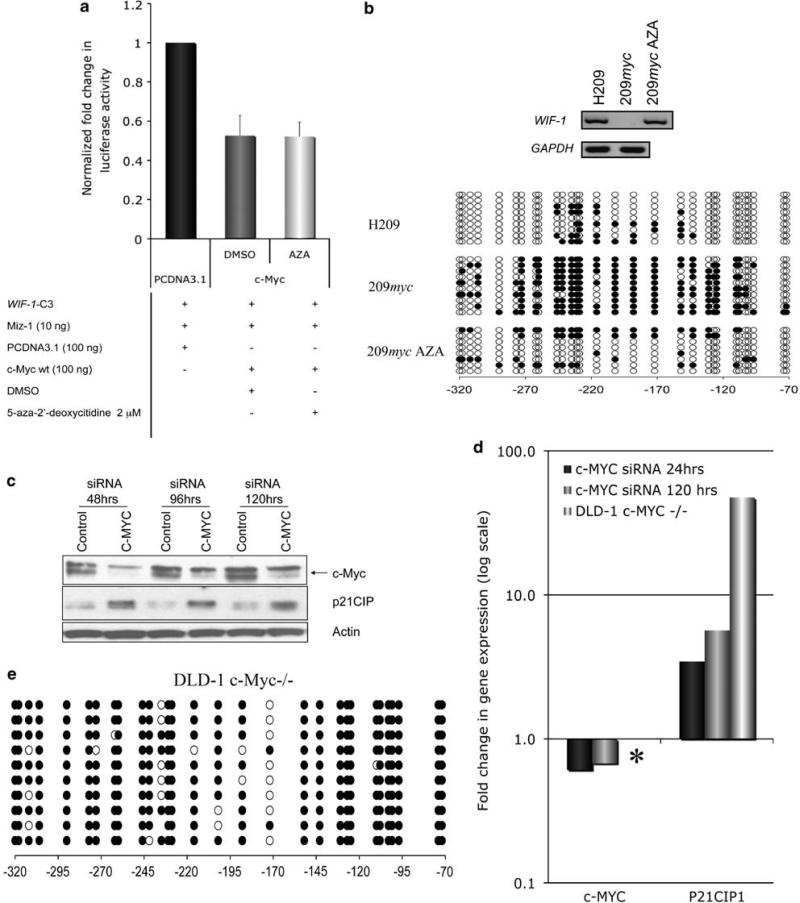
The Wnt signaling pathway is capable of self-regulation through positive and negative feedback mechanisms. For example, the oncoprotein c-Myc, which is upregulated by Wnt signaling activity, participates in a positive feedback loop of canonical Wnt signaling through repression of Wnt antagonists DKK1 and SFRP1. In this study, we investigated the mechanism of Wnt inhibitory factor-1 (WIF-1) silencing. Mapping of CpG island methylation of the WIF-1 promoter reveals regional methylation (-295 to -95 bp from the transcription start site) that correlates with transcriptional silencing. We identified Miz-1 as a direct activator of WIF-1 transcriptional activity, which is found at WIF-1 promoter. In addition, we show that c-Myc contributes to WIF-1 transcriptional repression in a Miz-1-dependent manner. Although the transient repression mediated by Miz-1/c-Myc is independent of de novo methylation, the stable repression by this complex is associated with CpG island methylation of the critical -295 to -95-bp region of the WIF-1 promoter. Importantly, Miz-1 and c-Myc are found at WIF-1 promoter in WIF-1 non-expressing cell lines DLD-1 and 209myc. Transient knockdown or somatic knockout of c-Myc in DLD-1 failed to restore WIF-1 expression suggesting that c-Myc is involved in initiating rather than maintaining WIF-1 epigenetic silencing. In a genome-wide screen, DNAJA4, TGFβ-induced and TRIM59 were repressed by c-Myc overexpression and DNA promoter hypermethylation. Our data reveal novel insights into c-Myc-mediated DNA methylation-dependent transcriptional silencing, a mechanism that might contribute to the dysregulation of Wnt signaling in cancer.


Figures

Figure 1
Figure 1
WIF-1 promoter methylation status. (a) Upper panel: WIF-1 expression was analyzed by RT–PCR in lung and colorectal cancer cell lines either untreated, treated with dimethylsulphoxide (DMSO) (m) or with 2 μm of 5-aza-2′-azadeoxycitidine treatment for 56 h (A56), 82 h (A82) or 96 h (A96). Glyceraldehyde 3-phosphate dehydrogenase (GAPDH) was used as a control. Lower panel: real-time RT–PCR analysis of WIF-1 transcript in NSCLC cell lines. WIF-1 expression was highest in H920 followed by H1435, H1299, H1703, U1752, normal human bronchial epithelial (NHBE), H460, H157, A549 and H125 and absent in H1155, H1666, H1395 and H1993. (b, c) Genomic bisulfite sequencing analysis of WIF-1 promoter region (b) –21/+209 bp, (c, left panel) –401/–350 and (c, right panel) –320/–72 bp. White-filled circles represent unmethylated CGs and black-filled circle methylated CGs. All regions are relative to the TSS.
Figure 2
Figure 2
Characterization of WIF-1 proximal promoter. (a) Diagram showing the regions of WIF-1 promoter which were analyzed for their ability to induce luciferase activity (upper panel), or by genomic bisulfite sequencing (lower panel). Vertical bars represent individual CGs. A candidate Sp1 binding site and a possible Inr sequence are also shown. (b) Luciferase activity of promoter deletion constructs, as shown in (b). pGL3-construct vector was used as a positive (POS), and empty pGL3-basic vector as a negative control (EV). (c) The promoter region –436 to +120 bp was in vitro methylated, cloned into pGL3 vector and luciferase activity was determined and compared with that of unmethylated WIF-1-C3. (d, e) Real-time ChIP was used to determine the enrichment of H3K4me2 (d) and H3K27me3 (e) at WIF-1 promoter (–836/–608 bp and –250/–50 bp) in H460, H1703 and H838. Enrichment was normalized to the value obtained for the H838 cell line (–836/–608 bp).
Figure 3
Figure 3
Miz-1 directly increases WIF-1 transcription. (a) Increasing amount of an empty vector (EV) or a plasmid encoding full-length Miz-1 were transiently transfected in 293 cells together with a luciferase construct driven by WIF-1 proximal promoter (WIF-1-C3) and pRL-TK. Luciferase values were normalized to renilla and are shown relative to empty vector. (b) Empty vector or a plasmid encoding full-length Miz-1 were transiently transfected in H1299 and after 24–48 h endogeneous WIF-1 transcript level was measured by Taqman RT–PCR (Miz-1 protein level is shown in the inset). Values for WIF-1 were normalized to a Tata box-binding protein (TBP) standard curve. (c) EV or a plasmid encoding full-length Miz-1 were transiently transfected in H1299 and after 24 samples were cross-linked with DSG and formaldehyde for ChIP analysis. Chromatin was immunoprecipitated with either a normal immunoglobulin G (IgG), Miz-1 or c-Myc antibodies and ChIP DNA was amplified using primers specific for WIF-1 promoter (region –50 to + 120 relative to the TSS). (d) SMARTpool ON-TARGETplus for Miz-1 (ZBTB17) was used to transiently knockdown Miz-1 level in H1299. Forty-eight hours following siRNA transfection, endogeneous WIF-1 transcript levels were measured by Taqman RT–PCR. Miz-1 protein levels were also determined (inset).
Figure 4
Figure 4
Miz-1/c-Myc co-operate to silence WIF-1 promoter activity. (a) In all, 100 ng of an empty vector (EV), c-Myc or c-Myc 394D were co-transfected with WIF-1-C3, pRL-TK together with or without 10 ng of plasmid encoding full-length Miz-1. At 48 h after transfection, luciferase and renilla was measured. Luciferase values were normalized to renilla and are relative to their corresponding empty vector. (b) ChIP was carried out with Miz-1, c-Myc or an IgG control in 209myc and DLD-1 cells were. ChIP DNA was amplified with primers specific for WIF-1 promoter (–50/+ 120 bp).
Figure 5
Figure 5
DNA methylation is involved in the stable but not transient repression of WIF-1. (a) The effect 5-aza-2′-deoxycitidine (48 h) or vehicle (dimethylsulphoxide (DMSO)) on c-Myc-mediated repression of Miz-1-driven WIF-1-C3 promoter activity was determined by luciferase assay. (b, upper panel) Expression of Wnt antagonist WIF-1 was determined by RT–PCR in H209, 209myc cells or 209myc cells treated with 5-aza-2′-deoxycitidine (209myc AZA) for 48 h. The expression of glyceraldehyde 3-phosphate dehydrogenase (GAPDH) was also monitored in these cell lines as a control. (b, lower panel) Genomic bisulfite sequencing was determined for WIF-1 promoter region –320/–72 bp. Other regions (–401/–350 bp; –21/+ 209 bp) of WIF-1 promoter were also analyzed (Supplementary Figure 3A and 3B). c-Myc depletion has no effect on WIF-1 promoter methylation status or expression level. (c) Western blot analysis of c-Myc after the transient treatment (48, 96 and 120 h) of colorectal cancer cell line DLD-1 with c-Myc SMARTpool siRNA. The expression of p21CIP1, a known target of c-Myc repression and the loading control β-actin was also analyzed. (d) Real-time RT–PCR was used to quantify c-MYC and P21CIP levels in wt DLD-1, DLD-1 –/– as well as DLD-1 cells transiently treated for 24 or 120 h with c-Myc siRNA. c-MYC was not detected in the knockout DLD-1 –/– cells (*). WIF-1 expression was not detected in either DLD-1 wt, DLD-1 c-Myc siRNA or c-Myc –/– (data not shown). Fold change in expression is relative to GAPDH. The graph is on a log scale. (e) Bisulfite genomic sequencing showing that WIF-1 promoter (–320/–72 bp) remains fully methylated in the knockout DLD-1 c-Myc –/– cells.
Figure 6
Figure 6
Identification of novel genes repressed by c-Myc and DNA promoter hypermetylation. (a) Scatter plot showing the fold change in expression in 209myc AZA relative to 209myc (Y axis) and H209 relative to 209myc cells (X axis). Each spot on the scatter plot represents one probe on the microarray. Probes for which the signal decreased because of c-Myc and increased as a result of 5-aza-2′-azadeoxycitidine treatment (fold change of at least 1.41) are located in the box on the top right hand side of the scatter plot. DNAJA4, TRIM59 and TGFβi are highlighted. (b, left panel) Real-time RT–PCR analysis of 36/185 CpG island-containing genes. RHO and BCAM were also included as controls (both genes were in the original list of 264-candidate genes) but have no CpG islands. Data are expressed as fold change in expression (log scale) relative to glyceraldehyde 3-phosphate dehydrogenase (GAPDH). (b, right panel) Gel-based RT–PCR shows that DNAJA4, TRIM59 and TGFβi were expressed in H209, silenced in 209myc and re-expressed in 209myc AZA. SP5, DKK3, ITM2C are control genes that showed only mild change in the real-time RT–PCR. GAPDH was used as a control. A negative control in which no reverse transcriptase was used in the cDNA synthesis is included (no RT).
Figure 7
Figure 7
Newly identified genes are silenced by DNA promoter hypermethylation. (a) Methylation-specific PCR (MSP) was used to assess the methylation status of DNAJA4, TRIM59 and TGFβi in cells lacking de novo methylation (DKO), an in vitro methylated DNA control (IVD), H209, 209myc and 209myc AZA as well as a water control. A signal in the U lane indicates an unmethylated promoter, a signal in the M lane identifies methylated promoter and a signal in both lanes suggests that the promoter is either hemimethylated or that a portion of the alleles are fully unmethylated while the rest is fully unmethylated. (b–d) Genomic bisulfite sequencing was employed to map out promoter methylation of CGs at DNAJA4, TRIM59 and TGFβi promoters in H209, 209myc and 209myc AZA cells. Arrows indicate the CGs, which were assessed by MSP analysis.

Similar articles

Cited by

References

    1. Ahmed AA, Mills AD, Ibrahim AE, Temple J, Blenkiron C, Vias M, et al. The extracellular matrix protein TGFBI induces microtubule stabilization and sensitizes ovarian cancers to paclitaxel. Cancer Cell. 2007;12:514–527. - PMC - PubMed
    1. Ai L, Tao Q, Zhong S, Fields CR, Kim WJ, Lee MW, et al. Inactivation of Wnt inhibitory factor-1 (WIF1) expression by epigenetic silencing is a common event in breast cancer. Carcino-genesis. 2006;27:1341–1348. - PubMed
    1. Barr LF, Campbell SE, Diette GB, Gabrielson EW, Kim S, Shim H, et al. c-Myc suppresses the tumorigenicity of lung cancer cells and down-regulates vascular endothelial growth factor expression. Cancer Res. 2000;60:143–149. - PubMed
    1. Benanti JA, Wang ML, Myers HE, Robinson KL, Grandori C, Galloway DA. Epigenetic down-regulation of ARF expression is a selection step in immortalization of human fibroblasts by c-Myc. Mol Cancer Res. 2007;5:1181–1189. - PubMed
    1. Bowen H, Biggs TE, Phillips E, Baker ST, Perry VH, Mann DA, et al. c-Myc represses and Miz-1 activates the murine natural resistance-associated protein 1 promoter. J Biol Chem. 2002;277:34997–35006. - PubMed

Publication types

MeSH terms

Substances