. 2010 Nov 4;29(44):5969-75.
doi: 10.1038/onc.2010.330. Epub 2010 Aug 9.

EMX2 is epigenetically silenced and suppresses growth in human lung cancer

Affiliations

EMX2 is epigenetically silenced and suppresses growth in human lung cancer

J Okamoto et al. Oncogene. .

Erratum in

  • Oncogene. 2010 Nov 4;29(44):5976. Beltran, A [corrected to Yagui-Beltran, A]

Abstract

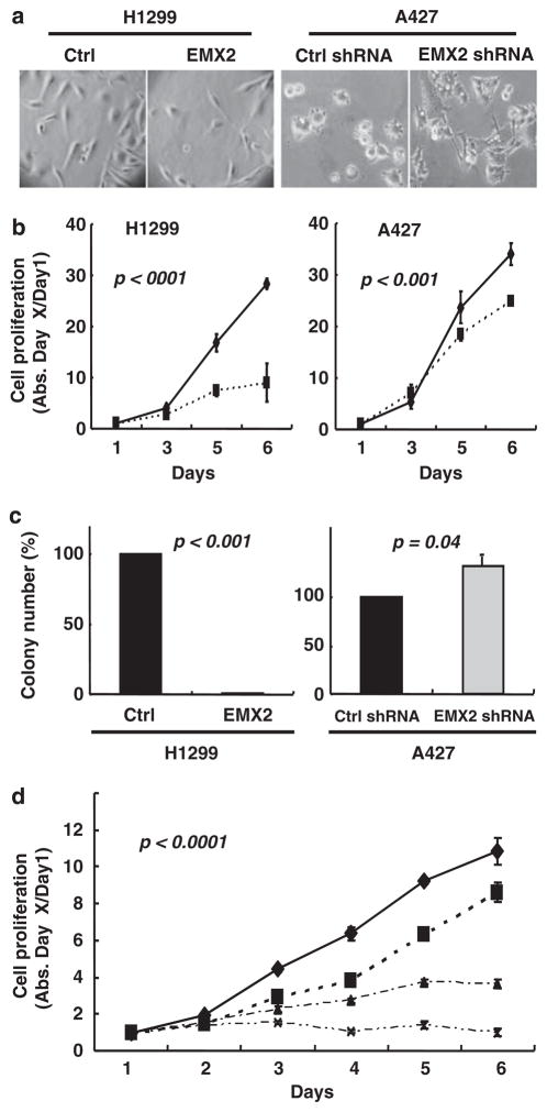
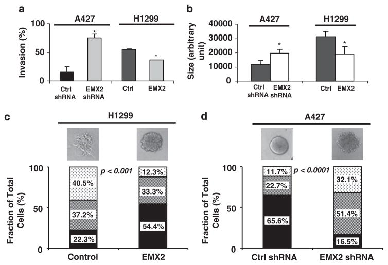
Lung cancer is a common cancer and the leading cause of cancer-related death worldwide. Aberrant activation of WNT signaling is implicated in lung carcinogenesis. EMX2, a human homologue of the Drosophila empty spiracles gene is a homeodomain-containing transcription factor. The function of EMX2 has been linked to the WNT signaling pathway during embryonic patterning in mice. However, little is known about the role of EMX2 in human tumorigenesis. In this study, we found that EMX2 was dramatically downregulated in lung cancer tissue samples and this downregulation was associated with methylation of the EMX2 promoter. Restoration of EMX2 expression in lung cancer cells lacking endogenous EMX2 expression suppressed cell proliferation and invasive phenotypes, inhibited canonical WNT signaling, and sensitized lung cancer cells to the treatment of the chemo cytotoxic drug cisplatin. On the other hand, knockdown of EMX2 expression in lung cancer cells expressing endogenous EMX2 promoted cell proliferation, invasive phenotypes and canonical WNT signaling. Taken together, our study suggests that EMX2 may have important roles as a novel suppressor in human lung cancer.


Conflict of interest statement

Conflict of interest

The authors declare no conflict of interest.

Figures

Figure 1
Figure 1
EMX2 expression was downregulated by methylation in lung cancer tissues and cell lines. Fresh samples (lung cancer tissue and its adjacent normal tissue) were collected from patients undergoing surgical resection with approval by the Committee on Human Research at the University of California, San Francisco (UCSF). Samples were promptly snap frozen in liquid nitrogen and stored at −170 °C before use. Total RNA was extracted using TRIzol LS (Invitrogen, Carlsbad, CA, USA). Human lung cancer cell lines were all purchased from American Type Culture Collection (ATCC, Manassas, VA, USA) and cultured in RPMI 1640 with 10% fetal bovine serum, penicillin (100 IU/ml)/streptomycin (100 μg/ml) at 37°C in a humidified 5% CO2 incubator. cDNA synthesis and Taqman PCR were performed as previously described (Raz et al., 2008). Hybridization probes and primers (Supplementary Information Table S1) were purchased from Applied Biosystems (ABI, Foster City, CA, USA). EMX2 expression of samples was calculated by using the 2−ddCt method (normalizing to their housekeeping gene GAPDH and then comparing to total RNA of adult normal lung tissue (BioChain, Hayward, CA, USA)). Quantitative methylation-specific PCR (qMSP) was performed as previously described (Fackler et al., 2004; Grote et al., 2005, 2006). Genomic DNA was extracted with Qiagen DNeasy kits (Qiagen, Valencia, CA, USA) and bisulfite modification of genomic DNA was performed using EZ DNA Methylation-Gold Kits (Zymo Research, Orange, CA, USA). Primers and probes (Supplementary Table S1) were designed using Primer Express and Methyl Primer Express Software v1.0 (ABI) and purchased from Operon (Huntsville, AL, USA). Relative EMX2 methylation levels were determined by using the 2−dCt method (normalizing to the housekeeping gene ACTB (Raz et al., 2008)) and then calculating the ratio (tumor/matched normal for tissues; cell line/an adult normal lung tissue (BioChain) for cell lines). Both quantitative RT–PCR and qMSP were done in triplicate using an ABI 7300 Real-time PCR System. (a) Quantitative RT–PCR of 64 tumors and their matched adjacent normal lung tissues. The y axis represents normalized relative EMX2 mRNA expression (arbitrary unit). (b) Quantitative RT–PCR (upper panel) and quantitative MSP (lower panel) of 10 representative tumors (black bars) compared with their matched adjacent normal lung tissues (gray bars). (c) Quantitative RT–PCR analysis and (d) Quantitative MSP analysis in lung cancer cell lines. An adult normal lung tissue was used as a control. Results are means±s.d. (error bars). (e) DAC treatment of lung cancer cell lines. Treatment of cells lines with 5μM DAC (Sigma, St Louis, MO, USA) was performed as previously described (Mazieres et al., 2004). Total RNA was isolated using Qiagen RNeasy kit 72 h after treatment, and EMX2 expression was examined by semiquantitative RT–PCR (primers (Supplementary Table S1) were purchased from Operon). GAPDH served as control for RNA quality and loading.
Figure 2
Figure 2
EMX2 suppressed lung cancer cell proliferation and sensitized lung cancer cells to cisplatin. H1299 cells were transfected with pcDNA 3.1/EMX2 mammalian expression vector (subcloned from pCMV6-XL5/EMX2 vector (Origene, Rockville, MD, USA)). A427 cells were transfected with EMX2 shRNAs (5′-TCAAGCCATTTACCAGGCTTCGGAGGAAG-3′ and 5′-CGG TGGAGAATCGCCACCAAGCAGGCGAG-3′) and non-silencing shRNA (all in pRFP-C-RS vector, Origene). Transfection was done using Lipofectamine2000 (Invitrogen). Transfected cells were re-plated from six-well plates to 10 cm dishes for selection with G418 (500 μg/ml; Invitrogen). Stable transfectants were maintained in regular medium with G418 (300 μg/ml) before analyses. (a) Morphology under light microscope (×40). (b) MTS assay of H1299 cells stably transfected with EMX2 (solid diamonds) and empty pCDNA3.1 vector control (solid squares); and A427 cells stably transfected with EMX2 shRNA (solid squares) and non-silencing shRNA control (solid diamonds). Controls were set as 100%. Proliferation assay was performed by plating the stably transfected cells in 96-well plates at a density of 500–1000 cells/well in 100 μl of G418 culture medium. Medium was changed every day. Cell viability was evaluated in triplicate by CellTiter 96 AQueous (Promega, Madison, WI, USA). (c) Colony formation assay. In all, 500 individual stably transfected cells were seeded in 10 cm dishes and cultured for 10 days. Colonies were then fixed by 10% formalin, stained with 0.5% crystal violet and counted. (d) Synergistic effect between EMX2 and cisplatin in H1299. Diamonds, squares, triangles and crosses are treatments of control vector alone, control vector+cisplatin (0.3 ng/ml), EMX2 cDNA alone and EMX2 cDNA+cisplatin (0.3 ng/ml), respectively. All results are means±s.d. (error bars). Differences between groups were compared with a two-sided Student’s t-test. A P-value of ≤0.05 was considered to be significant.
Figure 3
Figure 3
EMX2 suppressed invasive phenotypes of lung cancer cells. (a) Invasion assay using trans-well chamber with and without matrigel (BD BioCoat Matrigel Invasion Chamber, BD Biosciences, Lexington, KY, USA) was performed in triplicate for each stable transfectant according to the manufacturer’s protocol. Cells from five different fields of each insert membrane were counted under a light microscope (×40) and percent invasion was determined as follows: % invasion=(mean # of cells invading through matrigel insert membrane/mean # of cells migrating through control insert membrane without matrigel)× 100. (b, c) Analyses of 3D cultures of stably transfected cells. Eight-chambered culture slides (BD) were coated with 35 μl growth-factor reduced Cultrex Basement Membrane Extract (Trevigen, Gaithersburg, MD, USA) per well and left to solidify for 15 min. H1299 or A427 cells were treated with trypsin and resuspended in regular culture medium with serum. Cultrex was added to a total concentration of 2%, and 500 μl of the cell suspension was added to each chamber of the matrigel-coated slide. Medium was replaced every 2 days. After 1 week, 100+ acini were measured for size and graded for disruption. (b) Quantification of size of spheroids. (c) and (d) Phenotypes of spheroids (H1299 and A427) were categorized into three types (round (solid filling), asymmetric (dense dotted) and disrupted (sparse dotted)) and quantified. Representative phenotypes in each treatment were also shown. Symbol (*) in each graph represents statistical significance (P<0.05) by two-sided Student’s t-test.
Figure 4
Figure 4
EMX2 suppressed canonical WNT signaling in lung cancer cells. (a) Quantitative RT–PCR of EMX2 expression in cell lines stably transfected with control or EMX2 expression vector (in H1299) and with non-silencing control or EMX2-specific shRNA (in A427). H1703 served as an EMX2 expression level control. (b, c) TOP/FOP luciferase assays (performed 24 h after transfection as previously described in Clement et al., 2008) in H1299 cells stably transfected with EMX2 cDNA and in A427 cells stably transfected with EMX2 shRNA, respectively. (d) Western blotting of key canonical WNT downstream effector (cytosolic β-catenin; antibody from BD Biosciences) and target protein (Cyclin D1; antibody from Cell Signaling Technology, Danvers, MA, USA). β-Actin (antibody from Sigma) was used as protein control. Cytosolic proteins were prepared by using NE-PER Nuclear and Cytoplasmic Extraction Reagents (Pierce Biotechnology, Rockford, IL, USA) according to the manufacturer’s protocol. (e) The effect of DAC on key canonical WNT downstream effectors. Semiquantitative RT–PCR was used to confirm reactivation of the EMX2 expression after treatment of cells lines with 10 μM DAC. DAC treatment and semiquantitative RT–PCR were performed as described in Figure 1e. GAPDH served as control for RNA quality and loading. Western blotting of cytosolic β-catenin and Cyclin D1 was performed as described in Figure 4d. β-Actin was used as protein control. (f) Microarray profiling of lung cancer cell line H1299 stably transfected with EMX2 and empty vector control. Partial heatmap plot of hierarchical clustering including S100A4 and S100P is shown. Total RNA quality was assessed using a Pico Chip on an Agilent 2100 Bioanalyzer (Agilent, Santa Clara, CA, USA). RNA was amplified and labeled with Cy3-CTP or Cy5-CTP using the Agilent low RNA input fluorescent linear amplification kits following the manufacturer’s protocol. Labeled cRNA was assessed using the Nandrop ND-100, and equal amounts of Cy3- and Cy5-labeled target were hybridized to Agilent whole-human genome 44 K ink-jet arrays. Hybridization samples were randomized on the 3 × 44K format to correct any batch bias. Hybridizations were performed for 14 h according to the manufacturer’s protocol. Arrays were scanned using the Agilent microarray scanner and raw signal intensities were extracted with Feature Extraction v9.5 software (Agilent).

Similar articles

Cited by

References

    1. Abate-Shen C. Deregulated homeobox gene expression in cancer: cause or consequence? Nat Rev Cancer. 2002;10:777–785. - PubMed
    1. Akiri G, Cherian MM, Vijayakumar S, Liu G, Bafico A, Aaronson SA. Wnt pathway aberrations including autocrine Wnt activation occur at high frequency in human non-small-cell lung carcinoma. Oncogene. 2009;28:2163–2172. - PMC - PubMed
    1. American Cancer Society. Cancer facts and figures. 2008.
    1. Boersma CJ, Bloemen M, Hendriks JM, van Berkel EA, Olijve W, van Zoelen EJ. Homeobox proteins as signal transduction intermediates in regulation of NCAM expression by recombinant human bone morphogenetic protein-2 in osteoblast-like cells. Mol Cell Biol Res Commun. 1999;2:117–124. - PubMed
    1. Clement G, Guilleret I, He B, Yagui-Beltrán A, Lin YC, You L, et al. Epigenetic alteration of theWnt inhibitory factor-1 promoter occurs early in the carcinogenesis of Barrett’s esophagus. Cancer Sci. 2008;99:46–53. - PMC - PubMed

Publication types

MeSH terms

Substances