

Protease-activated receptor-2 mediates the expression of inflammatory cytokines, antimicrobial peptides, and matrix metalloproteinases in keratinocytes in response to Propionibacterium acnes
- PMID: 20697725
- PMCID: PMC2970807
- DOI: 10.1007/s00403-010-1074-z
Protease-activated receptor-2 mediates the expression of inflammatory cytokines, antimicrobial peptides, and matrix metalloproteinases in keratinocytes in response to Propionibacterium acnes
Abstract
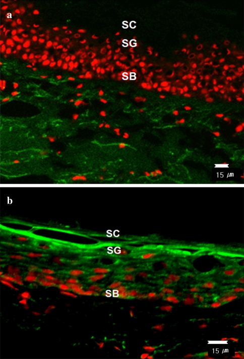
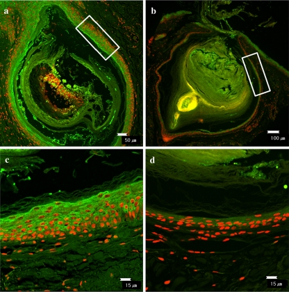
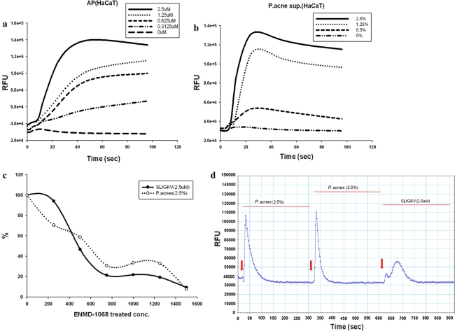
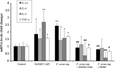
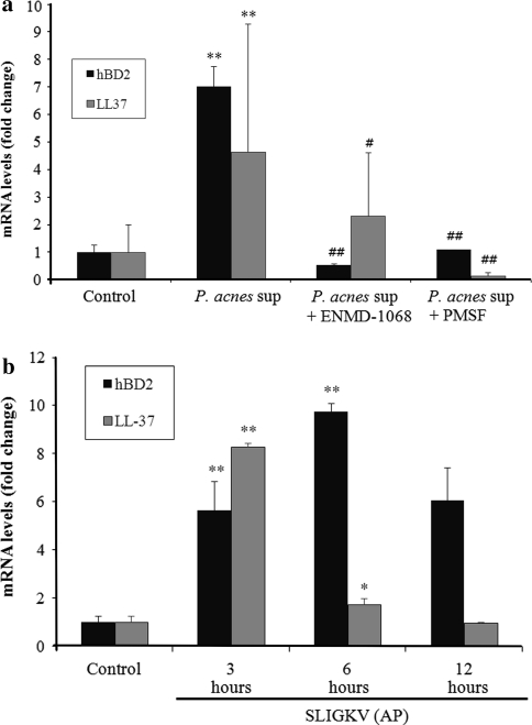
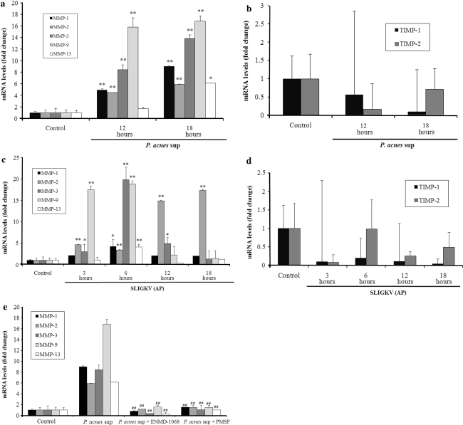
Propionibacterium acnes (P. acnes) has been known to produce various exogenous proteases, however, their role in acne pathogenesis remains largely unknown. Proteases elicit cellular responses, at least in part, via proteinase-activated receptor-2 (PAR-2), which is known to mediate inflammation and immune response. In this study, we investigated whether proteases from P. acnes could activate PAR-2 on keratinocytes and induce pro-inflammatory cytokines, antimicrobial peptides (AMPs), and matrix metalloproteinases (MMPs) via PAR-2 signaling. We examined PAR-2 expression and protease activity in acne lesions using immunofluorescence staining and in situ zymography. The effect of the culture supernatant of P. acnes on Ca(2+) signaling in immortalized keratinocytes (HaCaT) was measured using a fluorescence method. HaCaT cells were treated with P. acnes strain ATCC 6919 culture supernatant, with or without pretreatment with serine protease inhibitor or selective PAR-2 antagonist and the gene expression of pro-inflammatory cytokines, AMPs, and MMPs was detected using real-time reverse transcription-polymerase chain reaction. We found that the protease activity and PAR-2 expression were increased in acne lesions. The P. acnes culture supernatant induced calcium signaling in keratinocytes via PAR-2 and stimulated the mRNA expression of interleukin (IL)-1α, -8, tumor necrosis factor (TNF)-α, human beta defensin (hBD)-2, LL-37, MMP-1, -2, -3, -9, and -13 in keratinocytes, which was significantly inhibited by serine protease inhibitor as well as selective PAR-2 specific antagonist. These results indicate that PAR-2 plays an important role in the pathogenesis of acne by inducing inflammatory mediators in response to proteases secreted from P. acnes.
Figures
Similar articles
-
Anti-inflammatory properties of a new undecyl-rhamnoside (APRC11) against P. acnes.Arch Dermatol Res. 2011 Dec;303(10):707-13. doi: 10.1007/s00403-011-1147-7. Epub 2011 Apr 3. Arch Dermatol Res. 2011. PMID: 21461892
-
Expression of Protease-Activated Receptor-2 in SZ95 Sebocytes and its Role in Sebaceous Lipogenesis, Inflammation, and Innate Immunity.J Invest Dermatol. 2015 Sep;135(9):2219-2227. doi: 10.1038/jid.2015.151. Epub 2015 Apr 16. J Invest Dermatol. 2015. PMID: 25880702
-
Suppression of Propionibacterium acnes Infection and the Associated Inflammatory Response by the Antimicrobial Peptide P5 in Mice.PLoS One. 2015 Jul 21;10(7):e0132619. doi: 10.1371/journal.pone.0132619. eCollection 2015. PLoS One. 2015. PMID: 26197393 Free PMC article.
-
What is new in the pathophysiology of acne, an overview.J Eur Acad Dermatol Venereol. 2017 Sep;31 Suppl 5:8-12. doi: 10.1111/jdv.14374. J Eur Acad Dermatol Venereol. 2017. PMID: 28805938 Review.
-
Does inflammatory acne result from imbalance in the keratinocyte innate immune response?Microbes Infect. 2010 Dec;12(14-15):1085-90. doi: 10.1016/j.micinf.2010.07.015. Epub 2010 Aug 5. Microbes Infect. 2010. PMID: 20691803 Review.
Cited by
-
The Immunogenetics of Acne.Adv Exp Med Biol. 2022;1367:137-154. doi: 10.1007/978-3-030-92616-8_6. Adv Exp Med Biol. 2022. PMID: 35286695
-
Protease-Activated Receptor 2 in inflammatory skin disease: current evidence and future perspectives.Front Immunol. 2024 Sep 5;15:1448952. doi: 10.3389/fimmu.2024.1448952. eCollection 2024. Front Immunol. 2024. PMID: 39301020 Free PMC article. Review.
-
Protease Activated Receptor 1 and Its Ligands as Main Regulators of the Regeneration of Peripheral Nerves.Biomolecules. 2021 Nov 10;11(11):1668. doi: 10.3390/biom11111668. Biomolecules. 2021. PMID: 34827666 Free PMC article. Review.
-
The role of antimicrobial peptides in chronic inflammatory skin diseases.Postepy Dermatol Alergol. 2016 Feb;33(1):6-12. doi: 10.5114/pdia.2015.48066. Epub 2016 Feb 29. Postepy Dermatol Alergol. 2016. PMID: 26985172 Free PMC article. Review.
-
A Novel 3D Skin Explant Model to Study Anaerobic Bacterial Infection.Front Cell Infect Microbiol. 2017 Sep 14;7:404. doi: 10.3389/fcimb.2017.00404. eCollection 2017. Front Cell Infect Microbiol. 2017. PMID: 28959685 Free PMC article.
References
MeSH terms
Substances
LinkOut - more resources
Full Text Sources
Other Literature Sources
Medical
Molecular Biology Databases
Miscellaneous
