. 2010 Oct 1;9(10):4952-9.
doi: 10.1021/pr100202a.

Human serum processing and analysis methods for rapid and reproducible N-glycan mass profiling

Affiliations

Human serum processing and analysis methods for rapid and reproducible N-glycan mass profiling

Scott R Kronewitter et al. J Proteome Res. .

Abstract

Glycans constitute a new class of compounds for biomarker discovery. Glycosylation is a common post-translational modification and is often associated with transformation to malignancy. To analyze glycans, they are released from proteins, enriched, and measured with mass spectrometry. For biomarker discovery, repeatability at every step of the process is important. Locating and minimizing the process variability is key to establishing a robust platform stable enough for biomarker discovery. Understanding the variability of the measurement devices helps understand the variability associated with the chemical processing. This report explores the potential use of methods expediting the enzymatic release of glycans such as a microwave reactor and automation of the solid-phase extraction with a robotic liquid handler. The study employs matrix-assisted laser desorption/ionization-Fourier transform ion cyclotron resonance mass spectrometry but would be suitable with any mass spectrometry method. Methods for system-wide data analysis are examined because proper metrics for evaluating the performance of glycan sample preparation procedures are not well established.


Figures

Figure 1
Figure 1
Figure 1a. Sinusoidal response of microwave power (watt) and sample temperature (degree Celsius) under normal mode reactor operation and a 37°C set point. Figure 1b. PNGase F enzymatic digestion temperature profiles. The solid line represents the standard water bath incubation at 37°C. The 6W and 20W air-cooled microwave reactor temperature profiles are presented. The temperature curves reach their equilibrium temperature eight minutes into the run.
Figure 1
Figure 1
Figure 1a. Sinusoidal response of microwave power (watt) and sample temperature (degree Celsius) under normal mode reactor operation and a 37°C set point. Figure 1b. PNGase F enzymatic digestion temperature profiles. The solid line represents the standard water bath incubation at 37°C. The 6W and 20W air-cooled microwave reactor temperature profiles are presented. The temperature curves reach their equilibrium temperature eight minutes into the run.
Figure 2
Figure 2
This grid diagram represents the number of glycans detected that are unique and in common between sample Set B and sample Set C. 50 glycans were detected in both sets while 5 were only detected in Set B and 9 were only detected in Set C.
Figure 3
Figure 3
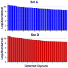
Specific glycan log-abundances from Set B and Set C are compared. Each bar represents a different glycan. Both plots are sorted by decreasing abundance.
Figure 4
Figure 4
Figure 4a. Replicate mass spectra from a single MALDI spot from the 10% ACN elution fraction. The relative distribution of the glycans is highly conserved between the technical replicate spectra. Annotated putative structures are depicted based common serum glycobiology. Figure 4b. Expanded view of low abundance glycans detected in technical replicate spectra from the same MALDI spot from the 10% ACN fraction. All ions detected were above the lower limit of detection set at 6 S/N.
Figure 4
Figure 4
Figure 4a. Replicate mass spectra from a single MALDI spot from the 10% ACN elution fraction. The relative distribution of the glycans is highly conserved between the technical replicate spectra. Annotated putative structures are depicted based common serum glycobiology. Figure 4b. Expanded view of low abundance glycans detected in technical replicate spectra from the same MALDI spot from the 10% ACN fraction. All ions detected were above the lower limit of detection set at 6 S/N.
Figure 5
Figure 5
Coefficient of variation vs. number of spectra collected within a spot. The variation decrease by roughly the square root of n where n is the number of replicate spectra averaged.
Figure 6
Figure 6
Coefficient of variation of each ion in the 10% ACN fraction vs. glycan mass. The plot is sorted by increasing variation. The coefficient of variation is calculated across a set of 8 MALDI spots from a single sample processed from Set C.
Figure 7
Figure 7
Frequency of detection from Set D. Frequency of detection is the fraction of samples of the sample set that contained the glycan above the noise level multiplied by one hundred.

References

    1. Apweiler R, Hermjakob H, Sharon N. On the frequency of protein glycosylation, as deduced from analysis of the SWISS-PROT database. Biochim Biophys Acta. 1999;1473(1):4–8. - PubMed
    1. Vance BA, Wu WY, Ribaudo RK, Segal DM, Kearse KP. Multiple dimeric forms of human CD69 result from differential addition of N-glycans to typical (Asn-X-Ser/Thr) and atypical (Asn-X-Cys) glycosylation motifs. Journal of Biological Chemistry. 1997;272(37):23117–23122. - PubMed
    1. Miletich JP, Broze GJ. Beta-Protein-C Is Not Glycosylated at Asparagine-329 - the Rate of Translation May Influence the Frequency of Usage at Asparagine-X-Cysteine Sites. Journal of Biological Chemistry. 1990;265(19):11397–11404. - PubMed
    1. Titani K, Kumar S, Takio K, Ericsson LH, Wade RD, Ashida K, Walsh KA, Chopek MW, Sadler JE, Fujikawa K. Amino-Acid-Sequences of Human Vonwillebrand-Factor. Biochemistry. 1986;25(11):3171–3184. - PubMed
    1. Lebrilla CB, An HJ. The prospects of glycan biomarkers for the diagnosis of diseases. Mol Biosyst. 2009;5(1):17–20. - PubMed

Publication types

MeSH terms