Self-confirming "AND" logic nanoparticles for fault-free MRI
- PMID: 20698661
- PMCID: PMC2935492
- DOI: 10.1021/ja104503g
Self-confirming "AND" logic nanoparticles for fault-free MRI
Abstract
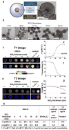
Achieving high accuracy in the imaging of biological targets is a challenging issue. For MRI, to enhance imaging accuracy, two different imaging modes with specific contrast agents are used; one is a T1 type for a "positive" MRI signal and the other is a T2 type for a "negative" signal. Conventional contrast agents respond only in a single imaging mode and frequently encounter ambiguities in the MR images. Here, we propose a "magnetically decoupled" core-shell design concept to develop a dual mode nanoparticle contrast agent (DMCA). This DMCA not only possesses superior MR contrast effects but also has the unique capability of displaying "AND" logic signals in both the T1 and T2 modes. The latter enables self-confirmation of images and leads to greater diagnostic accuracy. A variety of novel DMCAs are possible, and the use of DMCAs can potentially bring the accuracy of MR imaging of diseases to a higher level.
Figures


Similar articles
-
Redoxable heteronanocrystals functioning magnetic relaxation switch for activatable T1 and T2 dual-mode magnetic resonance imaging.Biomaterials. 2016 Sep;101:121-30. doi: 10.1016/j.biomaterials.2016.05.054. Epub 2016 Jun 1. Biomaterials. 2016. PMID: 27281684
-
Rational Design of Magnetic Nanoparticles as T1-T2 Dual-Mode MRI Contrast Agents.Molecules. 2024 Mar 18;29(6):1352. doi: 10.3390/molecules29061352. Molecules. 2024. PMID: 38542988 Free PMC article. Review.
-
An Ultrahigh-Field-Tailored T1 -T2 Dual-Mode MRI Contrast Agent for High-Performance Vascular Imaging.Adv Mater. 2021 Jan;33(2):e2004917. doi: 10.1002/adma.202004917. Epub 2020 Dec 2. Adv Mater. 2021. PMID: 33263204
-
Self-Confirming Magnetosomes for Tumor-Targeted T1 /T2 Dual-Mode MRI and MRI-Guided Photothermal Therapy.Adv Healthc Mater. 2022 Jul;11(14):e2200841. doi: 10.1002/adhm.202200841. Epub 2022 May 27. Adv Healthc Mater. 2022. PMID: 35579102
-
Inorganic nanoparticle-based T1 and T1/T2 magnetic resonance contrast probes.Nanoscale. 2012 Oct 21;4(20):6235-43. doi: 10.1039/c2nr31865b. Nanoscale. 2012. PMID: 22971876 Review.
Cited by
-
Development of hollow ferrogadolinium nanonetworks for dual-modal MRI guided cancer chemotherapy.RSC Adv. 2019 Jan 18;9(5):2559-2566. doi: 10.1039/c8ra09102a. eCollection 2019 Jan 18. RSC Adv. 2019. PMID: 35520519 Free PMC article.
-
Impact of metallic trace elements on relaxivities of iron-oxide contrast agents.RSC Adv. 2019 Sep 30;9(53):30932-30936. doi: 10.1039/c9ra07227f. eCollection 2019 Sep 26. RSC Adv. 2019. PMID: 35529357 Free PMC article.
-
Theranostic Calcium Phosphate Nanoparticles With Potential for Multimodal Imaging and Drug Delivery.Front Bioeng Biotechnol. 2019 Jun 4;7:126. doi: 10.3389/fbioe.2019.00126. eCollection 2019. Front Bioeng Biotechnol. 2019. PMID: 31214583 Free PMC article.
-
[T1-weighted magnetic resonance imaging contrast agents and their theranostic nanoprobes].Nan Fang Yi Ke Da Xue Xue Bao. 2020 Mar 30;40(3):427-444. doi: 10.12122/j.issn.1673-4254.2020.03.24. Nan Fang Yi Ke Da Xue Xue Bao. 2020. PMID: 32376585 Free PMC article. Review. Chinese.
-
Gd and Eu Co-Doped Nanoscale Metal-Organic Framework as a T1-T2 Dual-Modal Contrast Agent for Magnetic Resonance Imaging.Tomography. 2016 Sep;2(3):179-187. doi: 10.18383/j.tom.2016.00226. Tomography. 2016. PMID: 30042963 Free PMC article.
References
-
- Kircher MF, Mahmood U, King RS, Weissleder R, Josephson L. Cancer Res. 2003;63:8122–8125. - PubMed
- Nahrendorf M, Zhang H, Hembrador S, Panizzi P, Sosnovik DE, Aikawa E, Libby P, Swirski FK, Weissleder R. Circulation. 2008;117:379–387. - PMC - PubMed
- Cheon J, Lee JH. Acc Chem Res. 2008;41:1630–1640. - PubMed
- Gao JH, Gu HW, Xu B. Acc Chem Res. 2009;42:1097–1107. - PubMed
-
- Lauffer RE. Chem Rev. 1987;87:901–927.
- Taylor KML, Jin AWJ. Angew Chem Int Ed. 2008;47:7722–7725. - PubMed
-
- Na HB, Song IC, Hyeon T. Adv Mater. 2009;21:2133–2148.
-
- Kubaska S, Sahai DV, Saini S, Hahn PF, Halpern E. Clinic Radiol. 2001;56:410–415. - PubMed
Publication types
MeSH terms
Substances
Grants and funding
LinkOut - more resources
Full Text Sources
Other Literature Sources
Medical

