. 2011 Feb;20(2):243-57.
doi: 10.1089/scd.2010.0250. Epub 2010 Oct 12.

Human adipose-derived stromal cells stimulate autogenous skeletal repair via paracrine Hedgehog signaling with calvarial osteoblasts

Affiliations

Human adipose-derived stromal cells stimulate autogenous skeletal repair via paracrine Hedgehog signaling with calvarial osteoblasts

Benjamin Levi et al. Stem Cells Dev. 2011 Feb.

Abstract

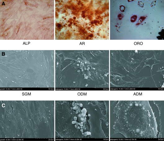
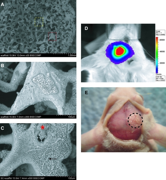
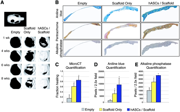
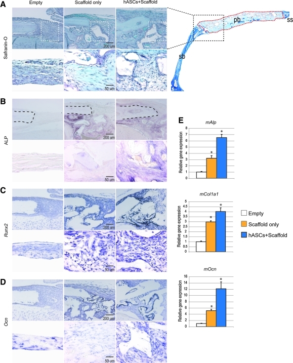
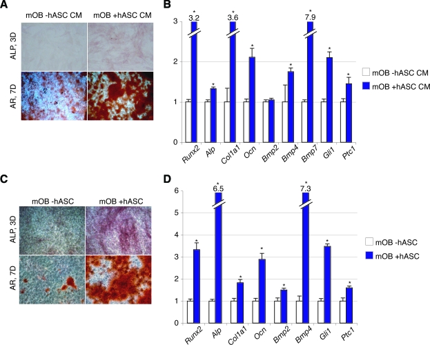
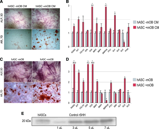
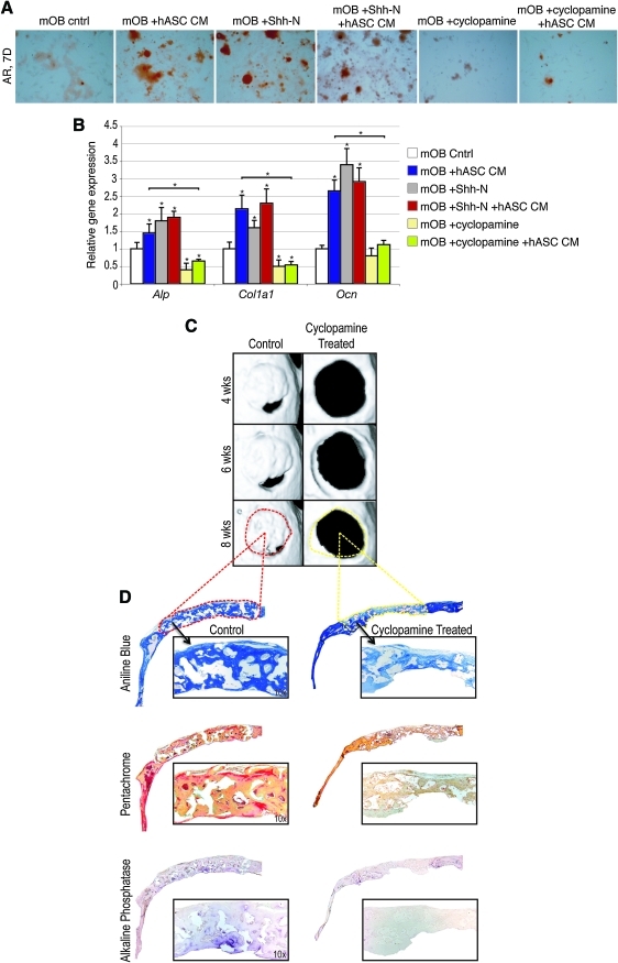
Human adipose-derived stromal cells (hASCs) have the proven capacity to ossify skeletal defects. The mechanisms whereby hASCs stimulate bone repair are not fully understood. In this study, we examined the potential for hASCs to stimulate autogenous repair of a mouse calvarial defect. Immunofluoresence, osteogenic stains, and surface electron microscopy were used to demonstrate osteogenic differentiation of hASCs. hASCs were engrafted into 4 mm calvarial defects in athymic mice using an osteoconductive scaffold. Analysis included microcomputed tomography, histology, in situ hybridization, and quantitative real-time-polymerase chain reaction. Next, the in vitro interaction between hASCs and mouse calvarial osteoblasts (mOBs) was assessed by the conditioned medium and coculture assays. The medium was supplemented with Hedgehog signaling modifiers, including recombinant N-terminal Sonic hedgehog, smoothened agonist, and cyclopamine. Finally, cyclopamine was delivered in vivo to hASC-engrafted defects. Significant calvarial healing was observed among hASC-engrafted defects compared with control groups (no treatment or scaffold alone) (*P<0.05). hASCs showed evidence of stimulation of host mouse osteogenesis, including (1) increased expression of bone markers at the defect edge by in situ hybridization, and (2) increased host osteogenic gene expression by species-specific quantitative real-time polymerase chain reaction. Using the conditioned medium or coculture assays, hASCs stimulated mOB osteogenic differentiation, accompanied by Hedgehog signaling activation. N-terminal Sonic hedgehog or smoothened agonist replicated, while cyclopamine reversed, the pro-osteogenic effect of the conditioned medium on mOBs. Finally, cyclopamine injection arrested bone formation in vivo. hASCs heal critical-sized mouse calvarial defects, this is, at least in part, via stimulation of autogenous healing of the host defect. Our studies suggest that hASC-derived Hedgehog signaling may play a paracrine role in skeletal repair.


Figures

FIG. 1.
FIG. 1.
Characterization of human adipose-derived stromal cells (hASCs). (A) Differentiation of hASCs toward osteoblast and adipocyte cell fates. (A, left) Alkaline phosphatase (ALP) staining after 3 days differentiation, demonstrating early osteoblast differentiation, 10×. (A, middle) Alizarin red (AR) staining after 7 days differentiation, indicating terminal osteoblast differentiation, 10×. (A, right) Oil red O (ORO) staining after 7 days adipogenic differentiation, showing intracellular lipid accumulation, 40×. (B, C) Surface electron microscopy of passage 1 hASCs after 3 days in the standard growth medium (SGM), osteogenic, or adipogenic differentiation medium (ODM or ADM). In SGM (left), hASCs have a smooth cell surface, while the appearance of bone nodules (center) or intracellular lipids (right) were appreciable with ODM and ADM, respectively, 600×–3,000× magnification. Color images available online at www.liebertonline.com/scd.
FIG. 2.
FIG. 2.
ASCs are engrafted and survive in a mouse cranial defect. (A) Appearance of hydroxyapatite-coated PLGA scaffold by surface electron microscopy, 35×. Higher magnification images of the areas within the dotted yellow and red lines can be seen in (B) and (C), respectively. (B) Scaffold appearance at 300×, hydroxyapatite appears coral-like with fine pore size. (C) hASCs attached on scaffold, 300×. Cells either spread out individually (black arrow) or remain in cell clusters (red asterisk). (D) Image of luciferase activity of a mouse 2 weeks postoperatively, after engraftment with Luc+ mouse ASCs. Scale on the right indicates intensity of signal, with red being the strongest signal. (E) Gross photographic image of nude CD-1 mouse 8 weeks after engraftment of hASCs. Encircled area shows the previous 4 mm parietal defect that was treated with a HA coated PLGA scaffold seeded with hASCs. Color images available online at www.liebertonline.com/scd.
FIG. 3.
FIG. 3.
hASCs heal mouse critical-sized defects. (A) Micro-computed tomography (CT) of defect sites postoperatively. Defects were either left empty or treated with scaffold only or hASCs with scaffold (n = 5 per group). Micro-CT images were obtained at stratified time points up to 8 weeks postoperatively. In upper left, a whole skull is provided for orientation. (B) After 8 weeks, mice were sacrificed for histological analysis. From left to right, defects shown include those left empty, treated with scaffold only, or treated with scaffold and hASCs. From top to bottom, staining includes aniline blue, pentachrome, and ALP, with bone appearing blue, yellow, and purple, respectively. (C) At 8 weeks, healing was quantified by micro-CT and Adobe Photoshop, expressed as average fraction osseous healing of the original defect size. (D) At 8 weeks, bone formation was quantified by Adobe Photoshop measurement of average aniline blue positive bone per 2.5 × field, n = 50 slides per group, 5 animals per group. (E) At 8 weeks, bone activity was quantified by Adobe Photoshop measurement of average ALP-positive bone per 2.5 × field, n = 5 slides per group, 5 animals per group. A one-factor analysis of variance (ANOVA) was utilized, followed by a post hoc Student's t-test to assess significance (*P < 0.05). Color images available online at www.liebertonline.com/scd.
FIG. 4.
FIG. 4.
hASCs stimulate mouse osteoblast (mOB) activity in a calvarial defect. (A) Histological analysis of the defect edge of defects left empty, treated with scaffold only, or treated with scaffold/hASCs at 2 weeks postoperatively. In the upper right corner, a low magnification image (2×) of the defect site in the right parietal bone (pb) and surrounding calvaria is shown for orientation purposes. The black dotted line depicts the edge of the defect site in the top panel of (A–D). The red dotted line outlines the defect site. Higher magnification images of the defect site, area outlined in white dotted lines in top panel of (A), can be seen in the bottom panel of (A–D). (A) Safranin-O staining, in which osteoid appears light blue. An absence of cartilage was noted in all sections, which would appear pink. (B) ALP staining, nonspecific for mouse versus human activity. (C) Runt-related transcription factor-2 (Runx2) expression by in situ hybridization, specific for mouse. (D) Osteocalcin (Ocn) expression by in situ hybridization, specific for mouse. (E) Quantitative real-time polymerase chain reaction analysis of RNA derived from cranial defects at 2 weeks postoperatively, specific for mouse genes. A significant induction of mAlp, mCol1a1, and mOcn was observed with hASC engraftment. n = 5 animals per group, *P < 0.05. A one-factor ANOVA was utilized, followed by a post hoc Student's t-test to assess significance. Sagittal suture (ss) and squamosal bone (sb). Color images available online at www.liebertonline.com/scd.
FIG. 5.
FIG. 5.
hASCs stimulate mOB differentiation in vitro. (A, B) mOBs were placed in osteogenic differentiation with or without supplementation of the hASC-conditioned medium (hASC CM). (A) ALP and AR staining of mOBs with or without hASC CM at 3 and 7 days differentiation, respectively. (B) Gene expression profile of mOBs after 3 days differentiation with or without hASC CM. Markers examined include osteoblast-specific genes, bone morphogenetic protein (BMP), and Hedgehog genes. (C, D) mOBs were place in osteogenic differentiation alone or in coculture of hASCs. (C) ALP and AR staining at 3 and 7 days, respectively, either alone or in coculture with hASCs. (D) Gene expression profile of mOBs after 3 days differentiation alone or in coculture with hASCs. n = 3 wells for all assays, *P < 0.05. A two-tailed Student's t-test was performed to assess significance. Color images available online at www.liebertonline.com/scd.
FIG. 6.
FIG. 6.
mOBs stimulate hASC differentiation in vitro. (A, B) hASCs were placed in ODM with or without supplementation of the mOB-conditioned medium (mOB CM). (A) ALP and AR staining of hASCs with or without mOB CM at 3 and 7 days differentiation, respectively. (B) Gene expression profile of hASCs after 3 days differentiation with or without mOB CM. Markers examined include osteoblast-specific genes, BMP, and Hedgehog genes. (C, D) hASCs were placed in osteogenic differentiation alone or in coculture of mOBs. (C) ALP and AR staining at 3 and 7 days, respectively, either alone or in coculture with mOBs. (D) Gene expression profile of hASCs after 3 days differentiation alone or in coculture with mOBs. n = 3 for all assays, *P < 0.05. A two-tailed Student's t-test was performed to assess significance. (E) Western immunoblot analysis of hASCs for Sonic hedgehog (SHH) demonstrating expression of the 20 kDa protein (left). This was run concurrently with different concentrations of recombinant human SHH as a positive control (right). Color images available online at www.liebertonline.com/scd.
FIG. 7.
FIG. 7.
Hedgehog signaling modulates the pro-osteogenic effect of hASC–mOB paracrine relationship. (A) mOBs were seeded in differentiation assays with N-terminal Sonic hedgehog (Shh-N) (250 ng/mL) or cyclopamine (20 mM) added to ODM, either alone or in combination with hASC CM. (A) AR staining at 7 days differentiation of mOBs under various conditions. (B) Osteogenic gene expression as assayed by quantitative real-time polymerase chain reaction at 3 days differentiation under various conditions. (C, D) Effects of cyclopamine administration to hASC-engrafted defects. A 50 μL suspension of cyclopamine (20 mM) was delivered by subcutaneous injection directly overlying the defect site for the first 3 postoperative days. (C) Micro-CT images were obtained at stratified time points up to 8 weeks postoperatively. Dotted red line represents area of histology for control below. Yellow dotted line depicts histology of cyclopamine-treated defect below. (D) Staining for aniline blue (above), pentachrome (middle), and ALP (below) was performed after 8 weeks healing. n = 3 for all assays, *P < 0.05. A 2-factor ANOVA was utilized (with or without hASC CM, with or without additional factors), followed by a post hoc Student's t-test to assess significance. Color images available online at www.liebertonline.com/scd.

Similar articles

Cited by

References

    1. HCUP. Agency for Healthcare Research and Quality; 2007. Healthcare cost and utilization project. - PubMed
    1. Fong KD. Nacamuli RP. Song HM. Warren SM. Lorenz HP. Longaker MT. New strategies for craniofacial repair and replacement: a brief review. J Craniofac Surg. 2003;14:333–339. - PubMed
    1. Nacamuli RP. Longaker MT. Bone induction in craniofacial defects. Orthod Craniofac Res. 2005;8:259–266. - PubMed
    1. Halvorsen YC. Wilkison WO. Gimble JM. Adipose-derived stromal cells—their utility and potential in bone formation. Int J Obes Relat Metab Disord. 2000;24(Suppl 4):S41–S44. - PubMed
    1. Lendeckel S. Jodicke A. Christophis P. Heidinger K. Wolff J. Fraser JK. Hedrick MH. Berthold L. Howaldt HP. Autologous stem cells (adipose) and fibrin glue used to treat widespread traumatic calvarial defects: case report. J Craniomaxillofac Surg. 2004;32:370–373. - PubMed

Publication types

MeSH terms

Substances

LinkOut - more resources