. 2010 Dec;161(7):1566-83.
doi: 10.1111/j.1476-5381.2010.00982.x.

Japanese encephalitis virus induces matrix metalloproteinase-9 in rat brain astrocytes via NF-κB signalling dependent on MAPKs and reactive oxygen species

Affiliations

Japanese encephalitis virus induces matrix metalloproteinase-9 in rat brain astrocytes via NF-κB signalling dependent on MAPKs and reactive oxygen species

Wei-Hsuan Tung et al. Br J Pharmacol. 2010 Dec.

Abstract

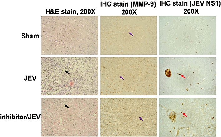
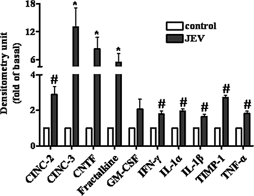
Background and purpose: Japanese encephalitis virus (JEV) is a member of the family Flaviviridae and JEV infection is a major cause of acute encephalopathy in children, which destroys cells in the CNS, including astrocytes and neurons. However, the detailed mechanisms underlying the inflammatory action of JEV are largely unclear.

Experimental approach: The effect of JEV on the expression of matrix metalloproteinase (MMP)-9 was determined by gelatin zymography, Western blot analysis, real-time PCR and promoter assay. The involvement of the NADPH oxidase and reactive oxygen species (ROS), MAPKs, and the transcription factor NF-κB in these responses was investigated by using selective pharmacological inhibitors and transfection with appropriate siRNAs.

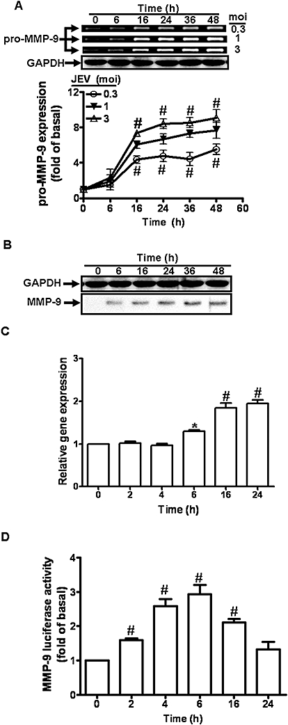
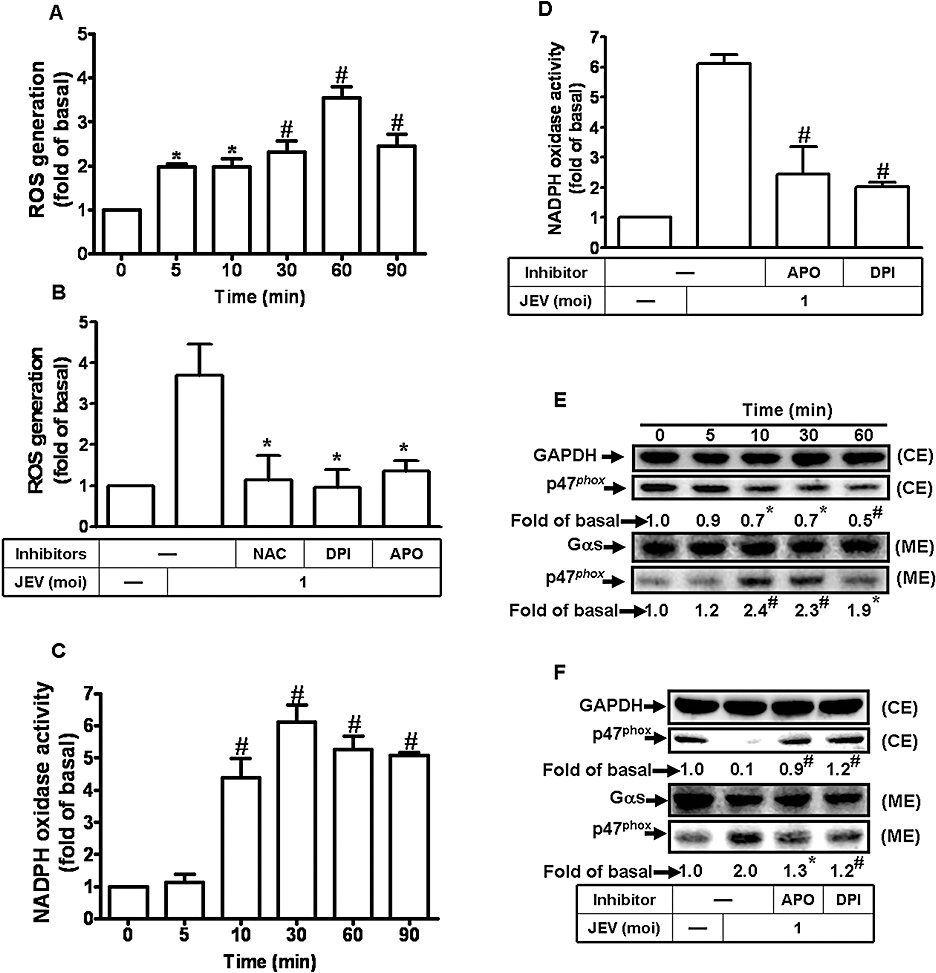
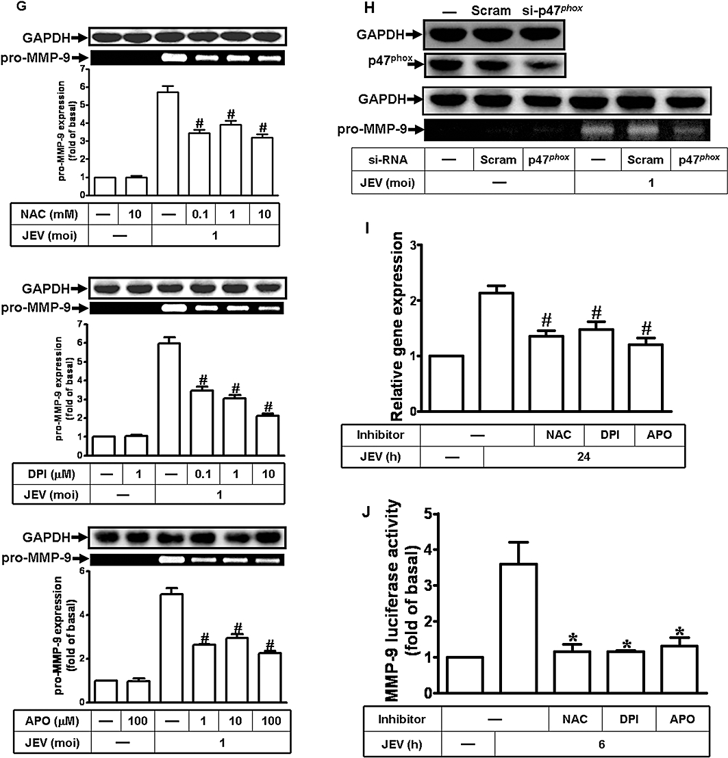
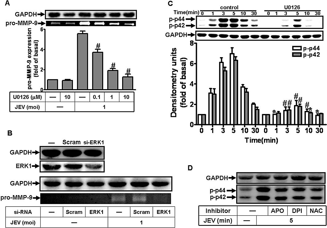
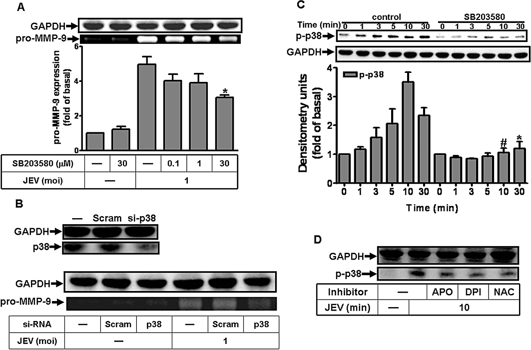
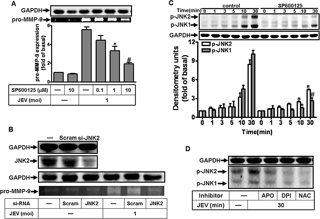
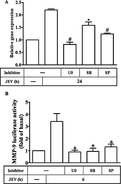
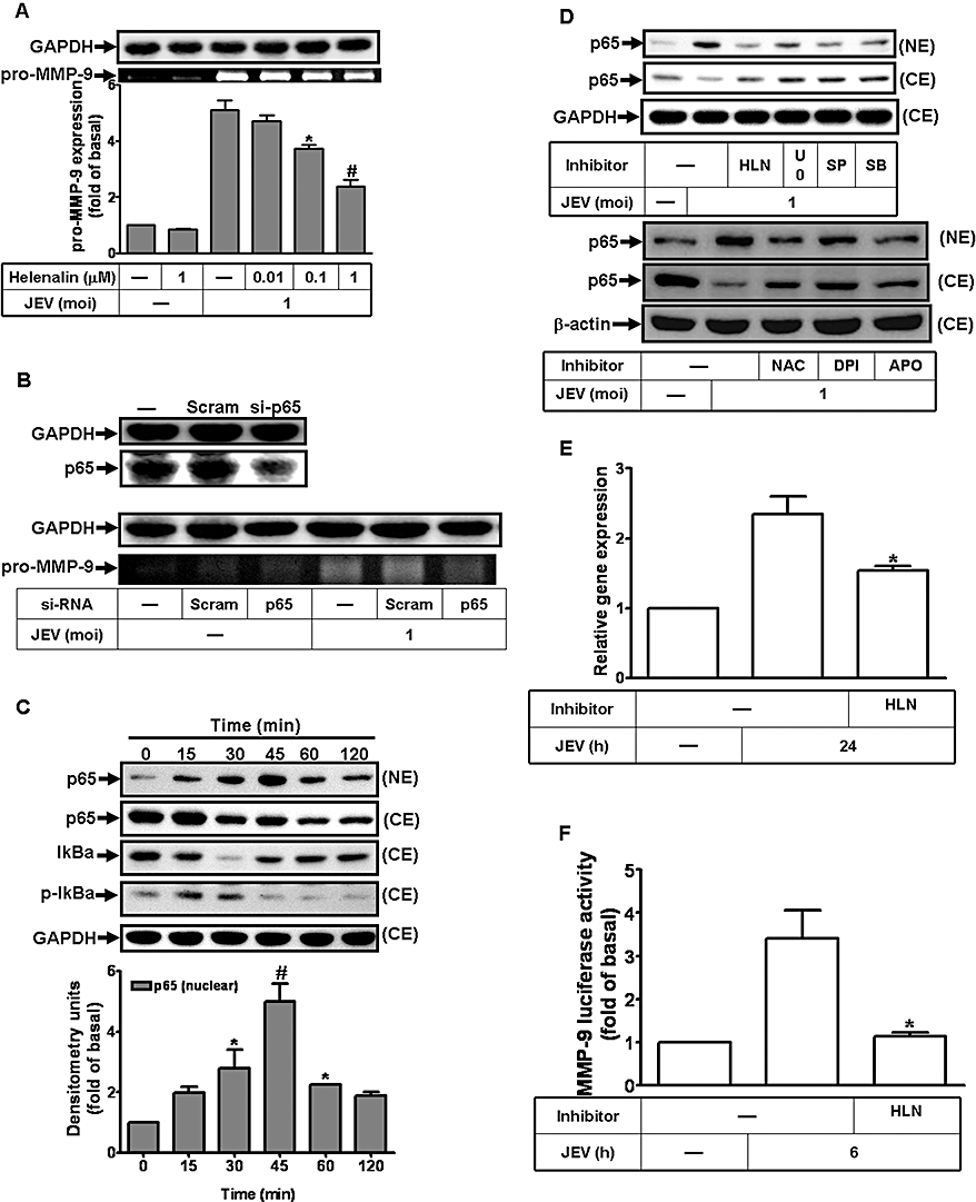
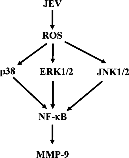
Key results: JEV induced the expression of the pro-form of MMP-9 in rat brain astrocytes (RBA-1 cells). In RBA-1 cells, JEV induced MMP-9 expression and promoter activity, which was inhibited by pretreatment with inhibitors of NADPH oxidase (diphenylene iodonium chloride or apocynin), MAPKs (U0126, SB203580 or SP600125) and a ROS scavenger (N-acetylcysteine), or transfection with siRNAs of p47(phox) , ERK1, JNK2 and p38. In addition, JEV-induced MMP-9 expression was reduced by pretreatment with an inhibitor of NF-κB (helenalin) or transfection with p65 siRNA. Moreover, JEV-stimulated NF-κB activation was inhibited by pretreatment with the inhibitors of NADPH oxidase and MAPKs.

Conclusions and implications: MMP-9 expression induced by JEV infection of RBA-1 cells was mediated through the generation of ROS and activation of p42/p44 MAPK, p38 MAPK and JNK1/2, leading to NF-κB activation.


Figures

Figure 1
Figure 1
Japanese encephalitis virus (JEV) induces matrix metalloproteinase (MMP)-9 expression in rat brain astrocytes (RBA)-1 cells. (A) RBA-1 cells were infected with JEV for various time intervals. The conditioned media were analysed by gelatin zymography. The whole cell lysates were analysed by Western blot using an anti-GAPDH antibody (as an internal control). (B) Cells were infected with JEV (moi = 1) for various time intervals. The conditioned media were analysed by Western blot using an anti-MMP-9 antibody. (C) RBA-1 cells were infected with JEV for various time intervals. The RNA samples were analysed by real-time PCR for the levels of MMP-9 mRNA. (D) Cells were transiently transfected with an MMP-9-luc reporter gene, and then infected with JEV for various time intervals. The promoter activity of MMP-9 was measured. Data are expressed as mean ± SEM of three independent experiments. *P < 0.05; #P < 0.01 as compared with the basal level.
Figure 2
Figure 2
Japanese encephalitis virus (JEV)-induced matrix metalloproteinase (MMP)-9 expression was mediated via the generation of reactive oxygen species (ROS) in rat brain astrocytes-1 cells. (A) Cells were labelled with DCF-DA (10 µM), and then infected with JEV (moi = 1) for the indicated time intervals. The fluorescence intensity (relative DCF fluorescence) was measured at 485 nm excitation and 530 nm emission. (B) Cells were labelled with DCF-DA, pretreated with 10 mM NAC, 10 µM diphenylene iodonium chloride (DPI) or 100 µM apocynin (APO) for 1 h, and then infected with JEV (moi = 1) for 1 h. The fluorescence intensity was measured. (C) Cells were infected with JEV (moi = 1) for the indicated time intervals. NADPH oxidase activity was measured. (D) Cells were pretreated with APO or DPI and then infected with JEV for 30 min. The NADPH oxidase activity was measured. (E) Cells were stimulated with JEV (moi = 1) for the indicated time intervals. The membrane and cytosolic fractions were analysed by Western blot using an anti-p47phox antibody. Gαs and GAPDH were used as marker proteins for membrane and cytosolic fractions respectively. (F) Cells were pretreated with DPI (10 µM) or APO (100 µM), and then infected with JEV (moi = 1) for 90 min. The membrane and cytosolic fractions were analysed by Western blot analysis using an anti-p47phox antibody. (G) Cells were pretreated with NAC, DPI or APO for 1 h, and then incubated with JEV (moi = 1) for 16 h. The level of MMP-9 expression was determined by gelatin zymography. (H) Cells were transfected with p47phox siRNA, and then treated with JEV for 16 h. The conditioned media were assessed for MMP-9 expression by gelatin zymography. The whole cell lysates were analysed by Western blot using an anti-p47phox or anti-GAPDH antibody. (I) Cells were pretreated with 10 mM NAC, 10 µM DPI or 100 µM APO for 1 h, and then incubated with JEV (moi = 1) for 6 h. The RNA samples were analysed by real-time PCR for the levels of MMP-9 mRNA. (J) Cells were transiently transfected with an MMP-9-luc reporter gene, pretreated with NAC, DPI or APO for 1 h, and then incubated with JEV for 6 h. The promoter activity of MMP-9 was measured. Data are expressed as mean ± SEM of three independent experiments. *P < 0.05; #P < 0.01 as compared with the basal level (A, C and E). *P < 0.05 as compared with the cells exposed to JEV alone (B, D, F, and J). CE, cystolic extract; ME, membranous extract.
Figure 2
Figure 2
Japanese encephalitis virus (JEV)-induced matrix metalloproteinase (MMP)-9 expression was mediated via the generation of reactive oxygen species (ROS) in rat brain astrocytes-1 cells. (A) Cells were labelled with DCF-DA (10 µM), and then infected with JEV (moi = 1) for the indicated time intervals. The fluorescence intensity (relative DCF fluorescence) was measured at 485 nm excitation and 530 nm emission. (B) Cells were labelled with DCF-DA, pretreated with 10 mM NAC, 10 µM diphenylene iodonium chloride (DPI) or 100 µM apocynin (APO) for 1 h, and then infected with JEV (moi = 1) for 1 h. The fluorescence intensity was measured. (C) Cells were infected with JEV (moi = 1) for the indicated time intervals. NADPH oxidase activity was measured. (D) Cells were pretreated with APO or DPI and then infected with JEV for 30 min. The NADPH oxidase activity was measured. (E) Cells were stimulated with JEV (moi = 1) for the indicated time intervals. The membrane and cytosolic fractions were analysed by Western blot using an anti-p47phox antibody. Gαs and GAPDH were used as marker proteins for membrane and cytosolic fractions respectively. (F) Cells were pretreated with DPI (10 µM) or APO (100 µM), and then infected with JEV (moi = 1) for 90 min. The membrane and cytosolic fractions were analysed by Western blot analysis using an anti-p47phox antibody. (G) Cells were pretreated with NAC, DPI or APO for 1 h, and then incubated with JEV (moi = 1) for 16 h. The level of MMP-9 expression was determined by gelatin zymography. (H) Cells were transfected with p47phox siRNA, and then treated with JEV for 16 h. The conditioned media were assessed for MMP-9 expression by gelatin zymography. The whole cell lysates were analysed by Western blot using an anti-p47phox or anti-GAPDH antibody. (I) Cells were pretreated with 10 mM NAC, 10 µM DPI or 100 µM APO for 1 h, and then incubated with JEV (moi = 1) for 6 h. The RNA samples were analysed by real-time PCR for the levels of MMP-9 mRNA. (J) Cells were transiently transfected with an MMP-9-luc reporter gene, pretreated with NAC, DPI or APO for 1 h, and then incubated with JEV for 6 h. The promoter activity of MMP-9 was measured. Data are expressed as mean ± SEM of three independent experiments. *P < 0.05; #P < 0.01 as compared with the basal level (A, C and E). *P < 0.05 as compared with the cells exposed to JEV alone (B, D, F, and J). CE, cystolic extract; ME, membranous extract.
Figure 3
Figure 3
Involvement of p42/p44 MAPK in Japanese encephalitis virus (JEV)-induced matrix metalloproteinase (MMP)-9 expression in rat brain astrocytes-1 cells. (A) Cells were pretreated with U0126 for 1 h and infected with JEV (moi = 1) for 16 h. (B) Cells were transfected with ERK1 siRNA, and then infected with JEV for 16 h. (A, B) The conditioned media were assessed for MMP-9 expression by gelatin zymography. The cell lysates were analysed by Western blot using an anti-ERK1 or anti-GAPDH antibody. (C) Cells were pretreated with 1 µM U0126 for 1 h and then infected with JEV (moi = 1) for the indicated time intervals. (D) Cells were pretreated with 10 mM NAC, 10 µM diphenylene iodonium chloride (DPI), or 100 µM apocynin (APO) for 1 h and then infected with JEV (moi = 1) for 5 min. The cell lysates were analysed by Western blot using an anti-phospho-p42/p44 MAPK or anti-GAPDH antibody. Data are expressed as mean ± SEM of three independent experiments. *P < 0.05; #P < 0.01 as compared with the cells exposed to JEV alone.
Figure 4
Figure 4
Involvement of p38 MAPK in Japanese encephalitis virus (JEV)-induced matrix metalloproteinase (MMP)-9 expression in rat brain astrocytes-1 cells. (A) Cells were pretreated with SB203580 for 1 h and then infected with JEV (moi = 1) for 16 h. (B) Cells were transfected with p38 siRNA, and then infected with JEV for 16 h. (A, B) The conditioned media were assessed for MMP-9 expression by gelatin zymography. The cell lysates were analysed by Western blot using an anti-p38 or anti-GAPDH antibody. (C) Cells were pretreated with 1 µM SB203580 for 1 h and then infected with JEV (moi = 1) for the indicated time intervals. (D) Cells were pretreated with 10 mM NAC, 10 µM diphenylene iodonium chloride (DPI), or 100 µM apocynin (APO) for 1 h and then infected with JEV (moi = 1) for 10 min. The cell lysates were analysed by Western blot using an anti-phospho-p38 MAPK or anti-GAPDH antibody. Data are expressed as mean ± SEM of three independent experiments. *P < 0.05; #P < 0.01 as compared with the cells exposed to JEV alone.
Figure 5
Figure 5
Involvement of JNK1/2 in Japanese encephalitis virus (JEV)-induced matrix metalloproteinase (MMP)-9 expression in rat brain astrocytes-1 cells. (A) Cells were pretreated with SP600125 for 1 h and then infected with JEV (moi = 1) for 16 h. (B) Cells were transfected with JNK2 siRNA, and then infected with JEV for 16 h. (A, B) The conditioned media were assessed for MMP-9 expression by gelatin zymography. The cell lysates were analysed by Western blot using an anti-JNK2 or anti-GAPDH antibody. (C) Cells were pretreated with 1 µM SP600125, 10 mM NAC, 10 µM diphenylene iodonium chloride (DPI) or 100 µM apocynin (APO) for 1 h, and then infected with JEV (moi = 1) for the indicated time intervals. (D) Cells were pretreated with 10 mM NAC, 10 µM DPI or 100 µM APO for 1 h, and then infected with JEV (moi = 1) for 30 min. The cell lysates were analysed by Western blot using an anti-phospho-JNK1/2 or anti-GAPDH antibody. Data are expressed as mean ± SEM of three independent experiments. *P < 0.05; #P < 0.01 as compared with the cells exposed to JEV alone.
Figure 6
Figure 6
Involvement of MAPKs in matrix metalloproteinase (MMP)-9 mRNA expression induced by Japanese encephalitis virus (JEV) in rat brain astrocytes-1 cells. (A) Cells were pretreated with 1 µM of U0126 (U0), SP600125 (SP) or SB203580 (SB) for 1 h, and then infected with JEV (moi = 1) for 6 h. The RNA samples were analysed by real-time PCR for the levels of MMP-9 mRNA. (B) Cells were transiently transfected with an MMP-9-luc reporter gene, pretreated with 1 µM of U0126, SB203580 or SP600125 for 1 h, and then infected with JEV for 6 h. The promoter activity of MMP-9 was measured. Data are expressed as mean ± SEM of three independent experiments. *P < 0.05; #P < 0.01 as compared with the cells exposed to JEV alone.
Figure 7
Figure 7
NF-κB translocation is a functional index during Japanese encephalitis virus (JEV)-induced matrix metalloproteinase (MMP)-9 expression in rat brain astrocytes-1 cells. (A) Cells were pretreated with helenalin (HLN) for 1 h and then infected with JEV (moi = 1) for 16 h. (B) Cells were transfected with p65 siRNA, and then infected with JEV for 16 h. (A, B) The conditioned media were assessed for MMP-9 expression by gelatin zymography. The cell lysates were analysed by Western blot using an anti-NF-κB p65 or anti-GAPDH antibody. (C, D) Time dependence of JEV-induced NF-κB translocation and IκB-α degradation. Cells were pretreated with or without 1 µM helenalin, 1 µM U0126, 1 µM SP600125, 1 µM SB203580, 10 mM NAC, 10 µM diphenylene iodonium chloride (DPI) and 100 µM apocynin (APO) for 1 h, and then were stimulated with JEV (moi = 1) for the indicated time intervals. Nuclear and cytosolic fractions were analysed by Western blot using an anti-NF-κB (p65), anti-phospho-IκBα, anti-IκBα antibody or anti-GAPDH antibody. (E) Cells were pretreated with 1 µM helenalin for 1 h, and then infected with JEV (moi = 1) for 6 h. The RNA samples were analysed by real-time PCR for the levels of MMP-9 mRNA. (F) Cells were transiently transfected with an MMP-9-luc reporter gene, pretreated with helenalin for 1 h and then infected with JEV for 6 h. The promoter activity of MMP-9 was measured. Data are expressed as mean ± SEM of three independent experiments. *P < 0.05; #P < 0.01 as compared with the cells exposed to JEV alone (A and F) or exposed to vehicle alone (C).
Figure 8
Figure 8
Japanese encephalitis virus (JEV) infection induced matrix metalloproteinase (MMP)-9 expression and brain damage in mice. Immunohistochemical staining (IHC) for MMP-9 and NS1 in sections of the brain and H&E staining for the brain from virus-free culture medium-treated mice (Sham), JEV-injected mice (JEV) and MMP-2/MMP-9 inhibitor II-pretreated mice (Inhibitor/JEV). The black arrow indicates tissue damage. The purple arrow indicates MMP-9 expression. The red arrow indicates NS1 glycoprotein of JEV. Photographs were taken at 200× magnification.
Figure 9
Figure 9
Cytokines released from rat brain astrocytes-1 cells induced by Japanese encephalitis virus (JEV) infection. After 24 h cultured in serum-free medium, the cells were treated with JEV (moi = 1) for 48 h. The cultured medium was collected and the levels of cytokines were determined using a cytokine array assay kit. Data are expressed as mean ± SEM of three independent experiments. *P < 0.05; #P < 0.01 as compared with the control.
Figure 10
Figure 10
Scheme of the JEV-mediated signalling pathways linked to matrix metalloproteinase (MMP-9) expression in brain astrocytes. Japanese encephalitis virus (JEV)-induced MMP-9 expression is mediated through NADPH oxidase/reactive oxygen species (ROS)/MAPKs leading to NF-κB activation. This signalling pathway might contribute to sustained expression of MMP-9 which is implicated in brain inflammation. Moreover, JEV may induce expression of some cytokines, including IL-1α, IL-β, TNF-α and IFN-γ in rat brain astrocytes-1 cells.

Similar articles

Cited by

References

    1. Arai K, Lee SR, Lo EH. Essential role for ERK mitogen-activated protein kinase in matrix metalloproteinase-9 regulation in rat cortical astrocytes. Glia. 2003;43:254–264. - PubMed
    1. Barber SA, Bruett L, Douglass BR, Herbst DS, Zink MC, Clements JE. Visna virus-induced activation of MAPK is required for virus replication and correlates with virus-induced neuropathology. J Virol. 2002;76:817–828. - PMC - PubMed
    1. Barbieri SS, Cavalca V, Eligini S, Brambilla M, Caiani A, Tremoli E, et al. Apocynin prevents cyclooxygenase 2 expression in human monocytes through NADPH oxidase and glutathione redox-dependent mechanisms. Free Radic Biol Med. 2004;37:156–165. - PubMed
    1. Behren A, Simon C, Schwab RM, Loetzsch E, Brodbeck S, Huber E, et al. Papillomavirus E2 protein induces expression of the matrix metalloproteinase-9 via the extracellular signal-regulated kinase/activator protein-1 signaling pathway. Cancer Res. 2005;65:11613–11621. - PubMed
    1. Bian ZM, Elner SG, Yoshida A, Kunkel SL, Su J, Elner VM. Activation of p38, ERK1/2 and NIK pathways is required for IL-1beta and TNF-alpha-induced chemokine expression in human retinal pigment epithelial cells. Exp Eye Res. 2001;73:111–121. - PubMed

Publication types

MeSH terms