. 2010 Aug 10:11:62.
doi: 10.1186/1471-2121-11-62.

Lamellipodium extension and membrane ruffling require different SNARE-mediated trafficking pathways

Affiliations

Lamellipodium extension and membrane ruffling require different SNARE-mediated trafficking pathways

Michael Skalski et al. BMC Cell Biol. .

Abstract

Background: Intracellular membrane traffic is an essential component of the membrane remodeling that supports lamellipodium extension during cell adhesion. The membrane trafficking pathways that contribute to cell adhesion have not been fully elucidated, but recent studies have implicated SNARE proteins. Here, the functions of several SNAREs (SNAP23, VAMP3, VAMP4 and syntaxin13) are characterized during the processes of cell spreading and membrane ruffling.

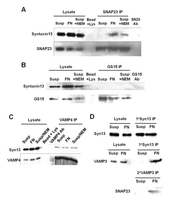
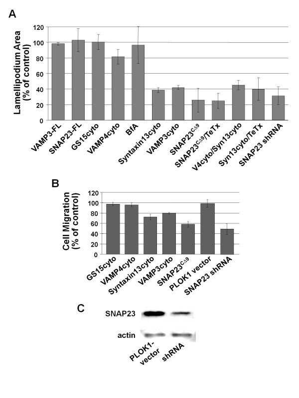
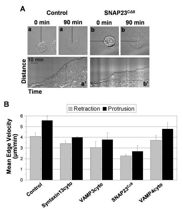
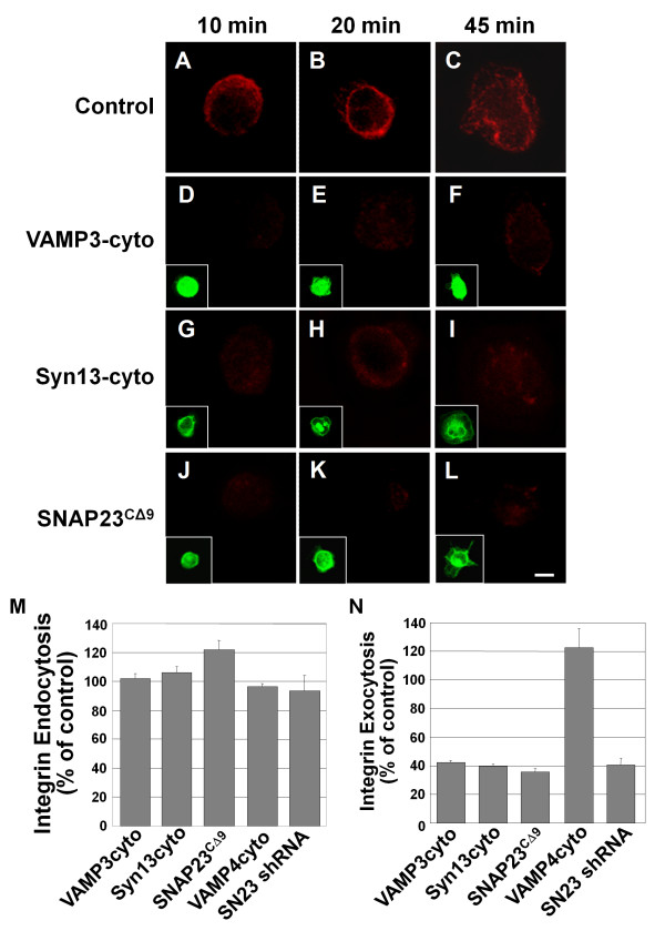
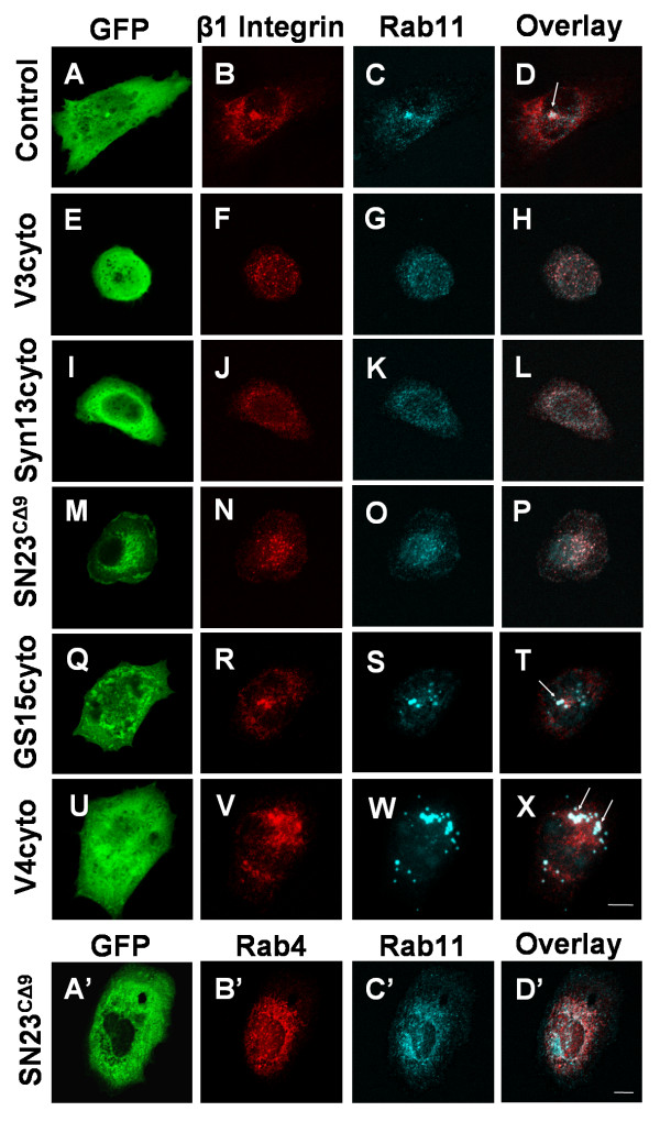
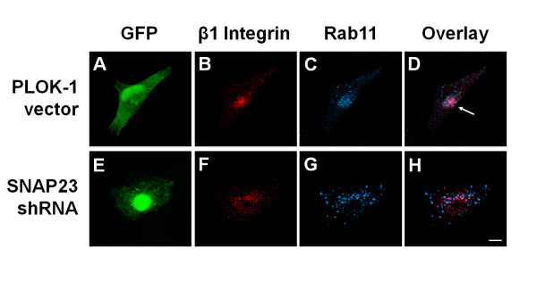
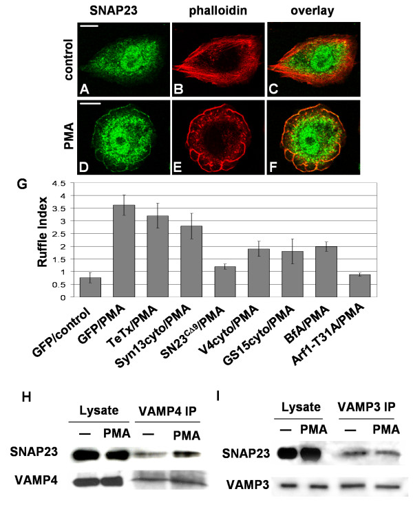
Results: We report the first description of a SNARE complex, containing SNAP23, syntaxin13 and cellubrevin/VAMP3, that is induced by cell adhesion to an extracellular matrix. Impairing the function of the SNAREs in the complex using inhibitory SNARE domains disrupted the recycling endosome, impeded delivery of integrins to the cell surface, and reduced haptotactic cell migration and spreading. Blocking SNAP23 also inhibited the formation of PMA-stimulated, F-actin-rich membrane ruffles; however, membrane ruffle formation was not significantly altered by inhibition of VAMP3 or syntaxin13. In contrast, membrane ruffling, and not cell spreading, was sensitive to inhibition of two SNAREs within the biosynthetic secretory pathway, GS15 and VAMP4. Consistent with this, formation of a complex containing VAMP4 and SNAP23 was enhanced by treatment of cells with PMA. The results reveal a requirement for the function of a SNAP23-syntaxin13-VAMP3 complex in the formation of lamellipodia during cell adhesion and of a VAMP4-SNAP23-containing complex during PMA-induced membrane ruffling.

Conclusions: Our findings suggest that different SNARE-mediated trafficking pathways support membrane remodeling during ECM-induced lamellipodium extension and PMA-induced ruffle formation, pointing to important mechanistic differences between these processes.


Figures

Figure 1
Figure 1
SNAP23-syntaxin13-VAMP3 interaction in CHO cells is stimulated by adhesion to FN. CHO-K1 cells were held in suspension after pretreatment with 1 mM NEM and 2 mM DTT (Susp), held in suspension after pretreatment with 1 mM NEM (Susp + NEM) or plated on FN (FN) for 20 min. The cells were lysed, and the indicated SNAREs were immunoprecipitated with antibodies coupled to sepharose beads from 1-1.5 mg of cellular protein. 10 μg aliquots of the lysates from which SNAREs were immunoprecipitated were run in 'Lysate' lanes. 'S13 Ab'/'SN23 Ab'/'GS15 Ab' indicate syntaxin13, SNAP23 and GS15 antibody control immunoprecipitations, where antibodies coupled to sepharose beads were incubated with lysis buffer, washed, and analyzed. (A) Syntaxin 13 immunoblot of SNAP23 immunoprecipitates showing immunoprecipitation of syntaxin 13 from cells plated on FN and cells treated with NEM, upper blot, and the same membrane reprobed for SNAP23, lower blot. (B) Syntaxin 13 immunoblot of GS15 immunoprecipitates, upper blot, and the same membrane reprobed for GS15, lower blot. (C) Syntaxin13 immunoblot of VAMP4 immunoprecipitates, and the same membrane reprobed for VAMP4, lower blot. (D) CHO-K1 cells, held in suspension (Susp) or plated on FN (FN), were lysed and syntaxin13 was immunoprecipitated with anti-syntaxin13 antibody. The syntaxin13 immunoprecipitates were washed and an aliquot (10%) of the eluate was analyzed by SDS-PAGE-western blot for syntaxin13, upper blot, and co-precipitated VAMP3, middle blot. The remainder of the syntaxin13-immunoprecipitate was re-suspended and immunoprecipitated with anti-VAMP3 antibody and then analyzed by SDS-PAGE-western blot for SNAP23, lower blot.
Figure 2
Figure 2
Dominant-negative SNARE domains impair lamellipodium extension. (A) CHO-K1 cells were transiently transfected with GFP-tagged constructs of the indicated SNAREs: GS15, VAMP4, VAMP3, syntaxin13 or SNAP23. Constructs encoded either the full length protein (FL) or the dominant-negative form (cyto; CΔ9 in the case of SNAP23). HeLa cells were transfected with a SNAP23 shRNA construct (SNAP23 shRNA). The cells were plated on FN-coated (20 μg/ml) coverslips and the extent of cell spreading was calculated as the increase in ventral surface area that occurred between 30 and 90 mins. The results are presented as percent of non-transfected control sample. More than 50 randomly selected transfected cells were analyzed per experiment. Means +/- SEM of at least three independent experiments are shown. (B) CHO cells were transfected as above, serum starved for 3 hrs and placed into a transwell chamber containing polycarbonate membranes coated on the underside with FN. Cells were allowed to migrate for 4.5 h, the membranes were fixed and the percentage of transfected cells migrated was assessed. Data is presented as the percentage of transfected cells migrated normalized to full-length controls and represent at least three independent experiments. (C) A sample of the same HeLa cells used in (A) were lysed and analyzed by Western blot for knockdown of SNAP23 expression. Actin served as a loading control. A set of representative blots are shown.
Figure 3
Figure 3
Inhibition of SNAREs reduces the rate of lamellipodium protrusion. Transfected CHO-K1 cells were plated in serum-free media on fibronetin for 90 min and monitored in real-time. (A) Representative control (a) and SNAP23CΔ9 (b) cells are shown at initial acquisition (0 min) and after 90 min (90 min) in the top panels. Kymographs (a' and b') in lower panels represent progression of the cell edge along the lines indicated with the cell edge highlighted. (B) The protrusion and retraction rate of the cell edge was determined from kymographs using ImageJ software. Data is presented as the mean velocity of immediate edge movement from at least two cell edges from at least 3 cells (when possible the most protrusive regions of the cell were measured).
Figure 4
Figure 4
Dominant-negative SNARE domains inhibit β1 integrin exocytosis. CHO-K1 cells were transiently transfected for 21 h with GFP-tagged constructs of truncated syntaxin13 (Syn13cyto)[D-F], VAMP3 (VAMP3cyto)[G-I], VAMP4 (VAMP4cyto) or SNAP23 (SNAP23CΔ9 - 15 h transfection) [J-L]. Untransfected control cells are shown in A-C. HeLa cells were transfected with a SNAP23 shRNA construct (SN23 shRNA) for 72 h. Cells were incubated with β1 integrin antibody in serum free media for 3 h to allowing internalization of the label. Then the cells were lifted with trypsin, removing remaining surface label, and were plated on 20 μg/mL FN for 10 min (A, D, G, J,), 20 min (B, E, H, K,) or 45 min (C, F, I, L). For exocytosis assays, cells were fixed and any surface exposed labeled-integrin was stained with AlexaFluor 594 secondary antibody (A-L, data for 20 min of adhesion shown in N). Insets show expression of GFP-tagged constructs (D-L). Images are 3 D reconstructions of a z-series. Scale bar represents 10 μm. (M and N) Integrin immunofluorescent staining at 20 min. was quantified in micrographs using ImageJ software. (M) To assess endocytosis of labeled integrin, cells were permeablized with 0.2% Trition X-100 in PBS before staining with AlexaFluor 594 secondary antibody (percent of non-transfected control). (N) Labeled integrin detected on the cell surface in non-permeabilized cells (percent of non-transfected control). Means ± SEM from three independent experiments (at least 20 cells per experiment) are shown in M and N.
Figure 5
Figure 5
Dominant-negative SNARE domains disrupt β1 integrin trafficking into the recycling endosome. CHO-K1 cells were transiently transfected for 22 h with GFP [A-D] or GFP-tagged constructs of truncated VAMP3 (V3cyto) [E-H], syntaxin13 (Syn13cyto) [I-L], SNAP23 (SNAP23CΔ9 - 15 h transfection) [M-P, A'-D'], GS15 (GS15cyto) [Q-T], or VAMP4 (V4cyto) [U-X]. Cells were labeled with anti-β1 integrin antibody in serum free medium for 1 h to allow internalization of integrin-antibody complexes. Cells were then washed in 0.2 M glycine, pH 2.5, fixed, and stained for Rab11 [C, G, K, O, S, W and C'] and the intracellular integrin-antibody complexes [B, F, J, N, R, and V], or Rab4 [B']. Overlays of the Rab11 and β1 integrin staining are shown in D, H, L, P, T and X, and Rab4 and Rab11 overlays are shown in D'. Arrows indicate conventional colocalization of β1 integrin in a central Rab11-containing compartment. Images are 3 D reconstructions of a z-series. Scale bar represents 10 μm.
Figure 6
Figure 6
shRNA-mediated downregulation of SNAP23 disrupts β1 integrin trafficking into the recycling endosome. Hela cells were transiently transfected for 72 h with GFP + PLOK-1 vectror [A-D] or GFP + vector containing SNAP23-targeting shRNA [E-H]. Cells were labeled with anti-β1 integrin antibody in serum free media for 1 h to allow internalization of integrin-antibody complexes. Cells were then washed in 0.2 M glycine, pH 2.5, fixed, and stained for the intracellular integrin-antibody complexes [B, F] and Rab11 [C, G]. Overlays of the Rab11 and β1 integrin staining are shown in D and H. Arrows indicate conventional colocalization of β1 integrin in a central Rab11-containing compartment. Images are 3 D reconstructions of a z-series. Scale bar represents 10 μm.
Figure 7
Figure 7
Analysis of SNARE function in PMA-induced membrane ruffle formation. CHO-K1 cells were treated with 500 nM PMA in DMEM (PMA) or DMEM alone (control) for 10 mins, fixed, permeabilized and stained with anti-SNAP23 antibody, Alexa-488-conjugated secondary antibody (A, D) and rhodamine-phalloidin (B, E). A-C, control cell; D-F, PMA-treated cell. Some cross-reactive binding of the SNAP23 antibody in the nucleus is observed (A, D). C and F are overlays of panels A and B and D and E, respectively. Scale bar represents 10 μm. (G) CHO-K1 cells were transfected with GFP vector alone, GFP-tagged dominant-negative constructs of the indicated SNAREs, or GFP together with the light chain of tetanus toxin. HeLa cells were transfected for 72 h with GFP + PLOK-1 or GFP + vector containing SNAP23-targeting shRNA. Cells were treated with 500 nM PMA in DMEM (PMA) or DMEM alone (control) for 10 mins, fixed, permeabilized and stained with rhodamine-phalloidin. Samples were then imaged by confocal microscopy and actin-rich ruffles on the dorsal region of the cells were quantified as described previously. Ruffle indices (expressed as mean +/- SEM) are shown from three independent experiments (more than 25 randomly selected transfected cells were analyzed per condition in each experiment). (H and I) CHO-K1 cells were treated as above, lysed, and VAMP4 (H) or VAMP3 (I) were immunoprecipitated. Immunoprecipitates were washed and analyzed by SDS-PAGE-western blotting for SNAP23, upper blots. Then membranes were stripped and reprobed for the precipitated SNARE, lower blots.

Similar articles

Cited by

References

    1. Nobes CD, Hall A. Rho, rac, and cdc42 GTPases regulate the assembly of multimolecular focal complexes associated with actin stress fibers, lamellipodia, and filopodia. Cell. 1995;81(1):53–62. doi: 10.1016/0092-8674(95)90370-4. - DOI - PubMed
    1. Sastry SK, Burridge K. Focal adhesions: a nexus for intracellular signaling and cytoskeletal dynamics. Exp Cell Res. 2000;261(1):25–36. doi: 10.1006/excr.2000.5043. - DOI - PubMed
    1. Powelka AM, Sun J, Li J, Gao M, Shaw LM, Sonnenberg A, Hsu VW. Stimulation-dependent recycling of integrin beta1 regulated by ARF6 and Rab11. Traffic. 2004;5(1):20–36. doi: 10.1111/j.1600-0854.2004.00150.x. - DOI - PubMed
    1. Sabe H. Requirement for Arf6 in cell adhesion, migration, and cancer cell invasion. J Biochem (Tokyo) 2003;134(4):485–489. - PubMed
    1. Roberts M, Barry S, Woods A, van der Sluijs P, Norman J. PDGF-regulated rab4-dependent recycling of alphavbeta3 integrin from early endosomes is necessary for cell adhesion and spreading. Curr Biol. 2001;11(18):1392–1402. doi: 10.1016/S0960-9822(01)00442-0. - DOI - PubMed

Publication types

LinkOut - more resources