Clinical Trial
. 2010 Aug 11:11:180.
doi: 10.1186/1471-2474-11-180.

Fluoroscopically guided transforaminal epidural dry needling for lumbar spinal stenosis using a specially designed needle

Affiliations
Clinical Trial

Fluoroscopically guided transforaminal epidural dry needling for lumbar spinal stenosis using a specially designed needle

Kang Ahn et al. BMC Musculoskelet Disord. .

Abstract

Background: This report describes the methodological approach and clinical application of a minimally invasive intervention to treat lumbar spinal stenosis (LSS).

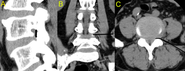
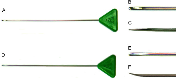
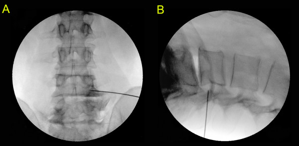
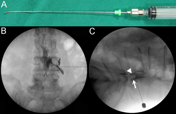
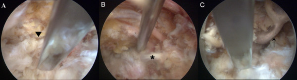
Methods: Thirty-four patients with LSS underwent fluoroscopically guided transforaminal epidural dry needling using a specially designed flexed Round Needle. The needle was inserted 8-12 cm lateral to the midline at the level of the stenosis and advanced to a position between the anterior side of the facet joint and pedicle up to the outer-third of the pedicle. The needle was advanced medially and backed laterally within a few millimetres along the canal side of the inferior articular process between the facet joint and pedicle. The procedure was completed when a marked reduction in resistance was felt at the tip of the needle. The procedure was performed bilaterally at the level of the stenosis.

Results: The average follow-up period was 12.9 +/- 1.1 months. The visual analogue scale (VAS) pain score was reduced from 7.3 +/- 2.0 to 4.6 +/- 2.5 points, the Oswestry Disability Index (ODI) score decreased from 41.4 +/- 17.2 to 25.5 +/- 12.6% and the average self-rated improvement was 52.6 +/- 33.1%. The VAS scores indicated that 14 (41.2%) patients reported a "good" to "excellent" treatment response, while 11 (32.4%) had a "good" to "excellent" treatment response on the ODI and 22 (64.7%) had a "good" to "excellent" treatment response on the self-rated improvement scale.

Conclusions: These results suggest that fluoroscopically guided transforaminal epidural dry needling is effective for managing LSS.


Figures

Figure 1
Figure 1
Three-dimensional CT images of the transforaminal epidural dry needling procedure. The sagittal CT image shows the area where the transforaminal epidural dry needling was performed (A). The coronal (B) and axial (C) CT images show the depth and route of needle insertion.
Figure 2
Figure 2
The needle used in fluoroscopically guided transforaminal epidural dry needling to treat lumbar spinal stenosis. The Round Needle before bending (A) and close-ups of its tip from above (B) and the side (C). The flexed Round Needle after bending (D) and close-ups of its tip from above (E) and the side (F).
Figure 3
Figure 3
Fluoroscopy images of the transforaminal epidural dry needling procedure. The anteroposterior (A) and lateral (B) fluoroscopy images are from a patient undergoing the intervention.
Figure 4
Figure 4
Transforaminal epidural contrast injection test to demonstrate that the specially designed needle effectively reached the target structures. The curved spinal injection needle used in the test (A). The anteroposterior fluoroscopy image shows a needle that has been advanced transforaminally at the L4-L5 level in the same manner as that used in the treatment and contrast agent flowing through the epidural space (B). The lateral fluoroscopy image. The triangle (▼) indicates contrast agent in the anterior epidural space when the needle was used in its original straight form. The arrow (↑) indicates contrast agent in the posterior epidural space when the curved needle was used. The curved needle contacted the canal side of the inferior articular process between the facet joint and pedicle (C).
Figure 5
Figure 5
A cadaveric examination used to evaluate the safety of fluoroscopically guided transforaminal epidural dry needling. The flexed Round Needle contacted and passed by the facet joint (▼) is advanced into the intervertebral foramen (A). The needle passes over the posterior longitudinal ligament (*) and reaches the anterior epidural space in the same manner as with the anterior epidural approach (B). After removing the posterior longitudinal ligament and dura mater by cauterisation, the tip of the needle is found to be located under the thecal sac (↑) without contacting it (C).

References

    1. Fritz JM, Delitto A, Welch WC, Erhard RE. Lumbar spinal stenosis: a review of current concepts in evaluation, management, and outcome measurements. Arch Phys Med Rehabil. 1998;79:700–708. doi: 10.1016/S0003-9993(98)90048-X. - DOI - PubMed
    1. Weinstein PR. Diagnosis and management of lumbar spinal stenosis. Clin Neurosurg. 1983;30:677–697. - PubMed
    1. Binder DK, Schmidt MH, Weinstein PR. Lumbar spinal stenosis. Semin Neurol. 2002;22:157–166. doi: 10.1055/s-2002-36539. - DOI - PubMed
    1. Katz JN, Harris MB. Clinical practice. Lumbar spinal stenosis. N Engl J Med. 2008;358:818–825. doi: 10.1056/NEJMcp0708097. - DOI - PubMed
    1. Elsig JP, Kaech DL. Imaging-based planning for spine surgery. Minim Invasive Ther Allied Technol. 2006;15:260–266. doi: 10.1080/13645700600958457. - DOI - PubMed

Publication types

MeSH terms